Assessment of DayÃÆâÃâââ¬Ãâââ¢s Methods for Critical Organs Doses Calculation of Breast Cancer Irradiation Compared with TLD
Mohammed A Ali Omer
1Department of Radiologic Technology, College of Applied Medical Science, Qassim University, Buraidah-KSA, Saudi Arabia
2College of Medical Radiologic Science, Sudan University of Science and Technology, Khartoum, Sudan
- *Corresponding Author:
- Mohammed A. Ali Omer
Department of Radiologic Technology
College of Applied Medical Science
Qassim University, P.O. Box 6800. Postal code 51452
Buraidah-KSA
Saudi Arabia
Tel: +966508037217
E-mail: alkajam@gmail.com
Received date: September 25, 2017; Accepted date: October 04, 2017; Published date: October 09, 2017
Citation: Omer MAA (2017) Assessment of Day’s Methods for Critical Organs Doses Calculation of Breast Cancer Irradiation Compared with TLD. Insights Med Phys. Vol. 2 No. 3:10
Abstract
The aim of this study was to assess the dose received by critical organs (eye, thyroid gland and the shoulder joint) in breast cancer irradiated patients using Day’s method technique and thermo-luminescence dosimeter TLD in addition to involved age group and cancer histological types. The study designed as experimental and retrospective study implies the location of critical organs, back scattered factor, source surface distance, patient’s ages and the histological cancer types. Excel data analysis revealed that: breast cancer has been observed among age groups of 18-23 and 24-29 years old and peaking at 30% among the age groups of 42-47 years old; the common histological types were ductal (57%), lobular (18%) and medullary (10%). The dose% received by critical organs decreased linearly and significantly (R2=0.6) by 5% cm-1, 1.3% cm-1 and 9.7% cm-1 (from supraclavicular) and 1.44% cm-1, 9.86% cm-1 and 1.83% cm-1 (from tangential field) respectively as the distance increase from the field boarder. Out of applied tumor dose (TD=4500 cGy); the critical organs received: 36, 319.5 and 382.5 cGy (Day’s method-anterior supraclavicular) respectively and 58.5, 355.5 and 436.5 cGy (TLD method). And from tangential field they received 13.5, 58.5 and 103 cGy (Day’s method) and 27, 99 and 135 cGy (TLD method). Day’s method generally showed only 0.6% less differs compared to TLD measurement.
Keywords
Breast; Radiotherapy; Critical organs; Day’s calculation; TLD
Introduction
The breast consists up of 15–20 lobules of glandular tissue embedded in fat; in mature woman it lies between the 2nd. Rib superiorly and extends to the infra-mammary fold at the level of the 6th-7th. Rrib in the vertical axis, horizontally it extends from the lateral edge of the sternum to the anterior or mid-axillary line. The posterior surface rests on the deep pectoralis fascia, serratus anterior, external oblique abdominal muscles, and the upper rectus sheath. Breast tissue also projects into the axilla as the axillary tail of Spence [1]. As the lymphatic system represents the potential root of cancer spread and accordingly the fact stated by some scholars was that: the length to diameter ratio of axillary lymphadenopathy fall within less than 1.5 cm indicate the malignancy in addition to lymph nodes shape visualized by ultrasound [2,3].
Breast cancer is a worldwide disease resulting in many deaths; it represents the second most common cancer in the world, as reported by American Cancer Society that showed about 1.3 million American women are annually diagnosed with BC and about 0.5 million die from the malignancy [4], while Ravichandran and Al-Zahrani, Ravichandran et al. [5] investigated the incidence of female breast cancer in the Gulf Cooperation Council (GCC) countries in relation to the established reproductive factors. A total of 4480 breast cancer cases were diagnosed in women during 1998-2002 among GCC country nationals. However, in Saudi Arabia; the epidemiological studies carried out by Ravichandran et al. Al-Qahtani [6] in which they reported that: BCa incidence was 19.8% of all the female cancers detected in the Kingdom it was revealed that the BCa as the second most common malignancy in women in KSA [7]. An earlier report by Saudi National Cancer Registry [8] revealed an increasing proportion of BCa among women of different ages from 10.2% (2000) to 24.3%. Most cases have been presented at late stages for treatment in African countries as well as in Saudi Arabia and some other Arabian countries, which is due to the lack of awareness by women, accessibility to screening methods, and availability of African-based research findings that would influence decision making at the governmental level [9]. However in Sudan, breast cancer has been recorded from 2541 cases in 2000 to 6303 cases in 2010 [10,11]. Among such increasing rate; breast cancer reported as the first top case with an incidence rate of 25.1 per 100,000 as has been estimated by Amany et al. [12].
The efforts of national and international organization as well as the health care institutions have been continuing to manage and eradicating such universe endemic disease using different modalities; such as surgery, radiation therapy, chemotherapy, hormonal therapy and immune therapy [13]; and due to shortage of surgery to eradicate the microscopic residual cancer cells, other modalities have been inevitable for radical treatment. Among these models, conventional radiation therapy has been used in developing countries of Mideast and Africa utilizing 60Co-teletherapy machine and linear accelerator that resulted in good to excellent outcomes in 80% to 95% of cases after breast irradiation to total doses of 45-50.4 Gy in daily fractions of 1.8 to 2.0 Gy [14,15] to receive a dose of 95%-107% of the prescribed dose by the planning target volume [16-18], which accompanied with some serious consequences due to considerable radiation dose received by adjacent vital organs (sensitive) (eye’s lens, thyroid gland, lungs, and shoulder joint) that may induce lens’s cataract, hypo/hyperthyroidism, lung’s pneumonia/fibrosis, joint stiffness. The appearance of eye’s radiation sickening has been noted within 4-8 years post radiotherapy course relative to total dose as 20% response with a dose of 40 Gy, 50% with a dose of 50 Gy and 100% response with a dose of 57 Gy [19]. Also radiation retinopathy (loss of vision or visual acuity) has been noted within 5 years post radiotherapy [20]; consequently Monroe et al. [21] reported that: after < 50 Gy, there was 4% rate of retinopathy received by at least 25% of the globe with conventional fractionation and modern conformal techniques. Whereas Marks et al. and Mayo et al. [22,23] stated that: “the whole organ dose of 50 Gy is associated with < 1% risk of blindness and between 55 and 60 Gy, the risk of blindness is approximately 3% to 7%). Regarding the joints response to radiation, a dose up to 6500 cGy in 2 Gy/fraction is tolerable, however the risk is proportional to the volume treated with doses greater than 55 Gy [24] such as radio-osteonecrosis which occur after 1-2 years of radiotherapy course among 2–20% of patients when irradiated with a fractionated radiation doses greater than 60-65 Gy [25]. The thyroid damage presented in forms of hypothyroidism, with low thyroxin and elevated thyroid stimulated hormone (TSH) has been noticed at a fractionated dose of >18 Gy [26] and the high risk occur after 5 years of irradiation or 8 years after fractionated irradiation at doses greater than 35 Gy which is less common [27,28]. Therefore the trend of this study will focus on the calculation of radiation dose received by these critical organs (eye, thyroid and shoulder joint) in external irradiation of breast cancer due to their presence near to radiation field using Day’s method calculation and TLD, which in turn serves the choices of implementing the best radiation technique and suiting the ethical issues in the field as well as to determine the accuracy of Day’s methods to be applicable and helpful for those countries who use conventional radiation therapy.
Methodology
This is an experimental study conducted in order to evaluate the dose received by critical organs in radical external breast irradiation in the radiation and Isotopes Center in Khartoum, in the period from May 2011 to March 2012.
Material
ÃÆâÃâââ¬âÃâê 60Cobalt teletherapy machine (1.25 MV) was used in the treatment of patient, with a dose rate (output) equal to 87.85 cGy/min and a percentage depth of 55% at 10 cm with a maximum depth dose (Dmax) at 0.5 cm. the tray factor=0.98 and a maximum field of 45 × 45 cm.
ÃÆâÃâââ¬âÃâê TLD crystal (LiF:Mg, Ti-Bicron NE, USA)
ÃÆâÃâââ¬âÃâê TLD reader (Harshaw model 3500, USA)
ÃÆâÃâââ¬âÃâê Ultrasound system Shimadzu-SBU 200 and probe (linear, 7.5-10 MHz)
ÃÆâÃâââ¬âÃâê An adult anthropomorphic phantom manufactured by CIRS (ATOM 701; CIRS, Norfolk, VA)
Method
The data of this study was collected from 153 patient underwent radiation therapy via four radiation fields (one anterior supraclavicle field, two tangential fields and posterior axilla field) encompassing the breast i.e. the bed of the tumor, internal mammary chain lymph nodes, cervical lymph nodes and axilla lymph nodes. And any patient irradiated via separate internal mammary chain field was excluded. The posterior axilla field considered when the finding revealed by ultrasound showed a length/diameter ratio of the lymph node was less than 1.5 cm in addition to the shape which was rendered the total sample as 110 patients.
Patient position: The patients’ position was on supine position on the slant board (with an appropriate angle) with her hand under her head and shoulder joint abducted at 90 degree. From such position; the following variables have been collected: field size, distance of the organ of interest from the field, treatment time, output, and backscatter factor, given dose, tumor dose and tray factor.
The distances of the critical organs eye, thyroid and shoulder joint from the anterior supra-clavicle and tangential fields then the % dose received by the critical were calculated using Day’s method [29,30] as follows: suppose ‘Q’ is a point outside the field at a distance ‘c’ cm from the field border as in Figures 1 and 2. Imagine a rectangle adjacent to the field such that it contains point ‘Q’ and has dimensions ‘2c-b’. Place another rectangle of dimensions a-b on the other side of ‘Q’ such that the field on the right of ‘Q’ is a mirror image of the field on the left, as shown in the figure. The dose at point Q at depth d is then given by subtracting the depth dose at ‘Q’ for field ‘2c × b’ from that; for field (2a+2c) × b and dividing by 2. The procedure is illustrated by the following example. Suppose it is required to determine percent depth dose at ‘Q’ (relative to Dmax at P) outside a (15 × 10) cm field at a distance of 5 cm from the field border. In diagram (1) then, a=15, b=10, and c=5. Suppose Q is at the center of the middle rectangle of dimensions 2c × b. Then the dose DQ at 10 cm depth is given by: ½ [DQ (40 × 10) - DQ (10 × 10)], If DQ is normalized to Dmax at P, one gets the percent depth dose at Q or %DQ as illustrated in Equation (1):
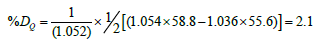

Figure 1: Shows the designing calculation of depth dose outside a rectangular radiation field based on Day’s method calculation of radiation dose outside of the irradiated field.

Figure 2: Shows the distribution of breast cancer based on the age groups in year.
Thus, as example for a 60C teletherapy machine beam at source surface distance (SSD)=80 cm the %DQ will be calculated as follows (Figure 1).
The doses received by the critical organs (eye, thyroid and shoulder joint) has been achieved from the irradiated breast phantom positioned typical to actual patients position with thermo-luminescence dosimeter (TLD) chips stapled outside the radiation fields at critical organs position which further read by TLD reader
(Harshaw model 3500, USA) and compared with Day’s method calculation to assess the accuracy of such method and recommend for applications in the field of radiation therapy.
Method of data analysis: The data was analyzed using Microsoft office excel under windows where the % dose for the point outside the field were calculated and verified by using an equation (2) that calculate the dose outside the filed using cosine theta (cos(θ)) , transmission factor and backscatter:
D = (output ×transmition×time×cos (θ )) / distance2 (2)
Then scatter plot between the Dose% received verse distances and field size were plotted with a regression line depicted the linear relationship between the two variable including the coefficient was obtained.
Results
The following section implies the figures for cancer distribution based on aging, types of cancer histopathology, percentage of radiation dose received by the eyes, thyroid and shoulder joint due to their position adjacent to the radiation fields of anterior supra-clavicle and tangential field.
Discussion and Analysis
The cancer has become as endemic fatal disease in Sudan recently with continuing high incidence as has been recorded from 2541 cases in 2000 to 6303 cases in 2010 [10,11]. Among such increasing rate; breast reported as the first top case with an incidence rate of 25.1 per 100,000 as has been estimated by Intisar et al. [12].
Based on the recent report about the high and continuing incidence of breast cancer in Sudan; a random selected sample consists of 150 patients showed that: breast cancer has been observed among the age groups of 18-23 and 24-29 years old with 7% and 11% respectively and peaking at 30% among the age groups of 42-47 years old then the incidence decrease rapidly after as shown in Figure 2; which is in a similar result stated by Northwestern University [31]. And the common histological types noted were the ductal, lobular and medullary with relative frequencies as 57%, 18% and 10% respectively (Figure 3). One notable fact was that: the incidence of breast cancer being predominated among the natives of Southern Sudan; such fact could be ascribed to inclusive social marriage in one tribe and as well inclusive among one family which still many tribes insisting to marry their daughters to only cousins’ sons and aunts’ sons leading to inheriting the hidden diseases. Also the environmental factors could play a major role in induction of cancer in such semi desert area of Sudan.

Figure 3: Shows the common histological types of breast cancer in the studied sample.
Based on this information and due to high incidence of patients’ complications (pneumonia, shoulder joint stiffness) during and/ or after radiotherapy course noted at radiation and isotopes center of Khartoum at radiation therapy section, the trend of assessing the radiation doses received by critical vital organs due to breast cancer irradiation could be appreciated.
As the radiation dose received by critical organs in external radiation therapy has been a matter of challenge up to date, and many techniques and methods have been adapted to calculate such dose. Day’s method has been used to calculate the dose received by critical organs (eye’s lens, thyroid gland and the shoulder joint) in breast cancer irradiation. Accordingly, and concise with inverse square law [30,32,33]; the dose % received by those critical organs have been decreased linearly and significantly (R2=0.6) as the distance in cm from the nearest field boarders increases (TLD calculation).
The dose % received by the eye relative to supraclavicular and tangential fields; decreased by a factor of 5% cm-1 and 1.31% cm-1 respectively. The dose % received by thyroid gland relative to supraclavicular and tangential fields decreased by a factor of 9.7% cm-1 and 1.44% cm-1 respectively, and the dose % received by the shoulder joint relative to supraclavicular and tangential fields decreased by a factor of 9.86% cm-1 and 1.83% cm-1 respectively. From which the analysis deduced that: closer critical organs to the boarder of the radiation field resulted in high exposure dose to organs; which ascribed to scattered radiation by collimator (40%) and head leakage [34,35] and due to penumbra profile at closer field boarder in 60Co-teletherapy machine that possess large radiation source size. On the other hand the increment of equivalent field size leads to increment of dose % received by the critical organs (Figure 4) in a linear significant trend (R2=0.5); in such relation and in case of anterior supraclavicular field (TLD methods) the eye, thyroid and shoulder joint received an average dose of (1.3, 7.9 and 9.7%) respectively and from Day’s method they received (0.8, 7.1 and 8.5%) respectively (Figure 5). And from tangential field (TLD method) the organs received (0.6, 2.2 and 3.0%) respectively and from Day’s Method they received (0.3, 1.3 and 2.3%) respectively (Figure 6).

Figure 4: Shows the correlation between the critical organ % dose and the square anterior supraclavicular field size in cm.

Figure 5: Shows the dose% received by critical organs measured by Day’s method and TLD in case of anterior supraclavicular fields.

Figure 6: Shows the dose% received by critical organs measured by Day’s method and TLD in case of tangential fields.
The high dose received by shoulder joint was due to it is vicinity to the radiation field and frequently inside the field which is being in needful to shielding.
And based on the prescribed total tumor dose (TD=4500–5000 cGy) for breast cancer [14,36] which is applicable in Sudan radiation therapy centers; the doses received by the (eye, thyroid gland, and shoulder joint) were: 36,319.5 and 382.5 cGy respectively (Anterior supraclavicular-Day’s method) and 58.5, 355.5 and 436.5 cGy respectively (anterior supraclavicular-TLD method) while from tangential field; the dose received by organs were: 13.5, 58.5, 103 cGy (Day’s method) and 27, 99 and 135 cGy (TLD method) respectively. The selection of therapeutic radiation dose range (4500-5000 cGy) consider as an attempt for avoiding the eye radiation reaction such as: ectropion, entropion with trichiasis and closure of the eyelid punctae (as chronic ones) and skin erythema and telangiectasia (acute ones) [37] and other may developed in other organs. In comparison with ICRP [38- 40], where they stated that: a dose of 3 Gy/1 fraction may cause cataract and the fractionated total body radiation therapy as (1.5- 2.0 Gy/fraction) to a dose of 12-14 Gy will induce a cataract risk by 10%, hence the results of current study deduced considerable incidence of lens cataract and other organs radiation sickening unless the therapeutic technologist utilizing trimmers of lead blocks, shield or the suitable techniques when implementing radiotherapy session for breast cancer.
In comparison between the dose received by critical organs outside the radiation field calculated by Day’s method and TLD versus the distance (Figure 7); Day’s method showed great success in estimation of radiation dose relative to measurement by TLD crystals and showed only 0.6% as less difference than TLD, such result would be helpful in radiation therapy for calculating the dose received by critical organs where TLD reader is unavailable.

Figure 7: Shows the correlation between the dose percent at specific organ measured by Day’s method and TLD versus the distance in cm of critical organ outside the radiation field.
Conclusion
Breast cancer has an increasing incidence rate among young Sudanese female and peaking among 42-47 years old. And the radiation therapy accompanied with considerable doses received by critical organs (eye, thyroid gland and shoulder joint). The dose received by critical organs outside the radiation field could be successfully assessed by Day’s method calculation; and it is utility could be maximized by simple computer programing to help those developing countries implementing the radiotherapy plan safely.
References
- Ellis H (2006) Clinical anatomy: A revision and applied anatomy for clinical students, (11th edn), Blackwell Publishing Ltd, USA.
- Esen G, Gurses B, Yilmaz MH, Ilvan S, Ulus S, et al. (2005) Gray scale and power doppler us in the preoperative evaluation of axillary metastases in breast cancer patients with no palpable lymph nodes. Eur Radiol 15: 1215-1223.
- Lernevall A (2000) Imaging of axillary lymph nodes. Acta Oncologica 39: 277-2781.
- American Cancer Society (2012) Cancer Facts and Figures, Atlanta: American Cancer Society, USA.
- Ravichandran K, Al-Zahrani AS (2009) Association of reproductive factors with the incidence of breast cancer in Gulf Cooperation Council countries. East Mediterr Health J 15: 612-621.
- Ravichandran K, Hamdan NA, Dyab AR (2005) Population based survival of female breast cancer cases in Riyadh Region, Saudi Arabia. Asian Pac J Cancer Prev 6: 72-76.
- Al-Qahtani MS (2007) Gut metastasis from breast carcinoma. Saudi Med J 28: 1590-1592.
- http: //www.scr.org.sa.
- Elgaili ME, Dafalla OA, Munazzah R, Arthur MM, Sulma IM (2010) Breast cancer burden in central Sudan. Int J Womens Health 2: 77-82.
- Mohammed EA, Alagib A, Babiker AI (2013) Incidents of cancer in Sudan: Past trends and future forecasts. Afr J Math 8: 136-142.
- Amany E, Muntaser EI, Dafalla A, Kamal Eldin HM, Sulma IM (2015) Part I: Cancer in Sudan burden, distribution, and trends breast, gynecological, and prostate cancers. Cancer Med 4: 447-456.
- Intisar ES, Hsin-Yi W, Kamal HM, Sulma IM (2014) Cancer incidence in Khartoum, Sudan: First results from the Cancer Registry, 2009-2010. Cancer Med 3: 1075-1084.
- Eric CE, Pointon RCS (1985) The Radiotherapy of malignant disease. Springer-Verlag Berlin Heidelberg New York Tokyo, Japan.
- James DC, Kian KA (2010) Radiation oncology: Rationale, technique, results. (9th edn), Elsevier Inc Philadelphia, USA. pp. 19103-2899.
- Wazer DE, DiPetrillo T, Schmidt-Ullrich R, Weld L, Smith TJ, et al. (1992) Factors influencing cosmetic outcome and complication risk after conservative surgery and radiotherapy for early-stage breast carcinoma. J Clin Oncol 10: 356-363.
- Magdalena PP, Julian M, Wojciech G (2012) Doses in organs at risk during head and neck radiotherapy using IMRT and 3D-CRT. Radiol Oncol 46: 328-336.
- Bhide SA, Nutting CM (2010) Advances in radiotherapy for head and neck cancer. Oral Oncol 46: 439-41.
- Grzadziel A, Grosu A, Kneschaurek P (2006) Three-dimensional conformal versus intensity-modulated radiotherapy dose planning in stereotactic radiotherapy: Application of standard quality parameters for plan evaluation. Int J Rad Oncol Biol Phys 66: S87-94.
- Leslie MD, Dische S (1994) The early changes in salivary gland function during and after radiotherapy given for head and neck cancer. Radiother Oncol 30: 26-32.
- Parsons JT, Bova FJ, Fitzgerald CR (1994) Radiation retinopathy after external beam irradiation: Analysis of time-dose factors. Int J Radiat Oncol Biol Phys 30: 765-773.
- Monroe AT, Bhandare N, Morris CG, Mendenhall WM (2005) Preventing radiation retinopathy with hyperfractionation. Int J Radiat Oncol Biol Phys 61: 856-864.
- Marks LB, Yorke ED, Jackson A (2010) Use of normal tissue complication probability models in the clinic. Int J Radiat Oncol Biol Phys 76: 10-19.
- Mayo C, Martel MK, Marks LB (2010). Radiation dosevolume effects of optic nerves and chiasm. Int J Radiat Oncol Biol Phys 76: S28-S35.
- Karasek K, Constine LS, Rosier R (1992) Sarcoma therapy: Functional outcome and relationship to treatment parameters. Int J Radiat Oncol Biol Phys 24: 651-656.
- Fajardo LF, Berthrong M, Anderson RE (2001) Radiation Pathology. Oxford University Press, New York, USA.
- Cohen LE (2005) Endocrine late effects of cancer treatment. Endocrinol Metab Clin N Am 34: 769-789.
- Hancock SL, Cox RS, McDougall IR (1991) Thyroid diseases after treatment of Hodgkin’s disease. N Engl J Med 325: 599-605.
- Sklar CA, Mertens AC, Walter A (2000) Changes in body mass index and prevalence of overweight in survivors of childhood acute lymphoblastic leukemia: Role of cranial irradiation. Med Pediatr Oncol 35: 91-95.
- Day M JA (1950) Note on the calculation of dose in x-ray fields. Br J Radiol 23: 368.
- Khan FM (2010) The physics of radiation therapy, (4th edn), Lippincott Williams & Wilkins, Philadelphia, PA 19106- USA.
- Eheman CR, Shaw KM, Ryerson AB, Miller JW, Ajani UA, et al. (2009) The changing incidence of in situ and invasive ductal and lobular breast carcinomas. Cancer Epidemiol Biomarkers Prev 18: 1763-1769.
- Northwestern University (2010) Radiation safety handbook. Office for research safety, Evanston, IL 60208, USA.
- Bernard S (1992) The health physics and radiological health handbook, Scinta, Inc, Silver Spring, MD 20902. USA.
- Michael LT, Tomas K (2011) Consideration of the radiation dose delivered away from the treatment field to patients in radiotherapy. J Med Phys 36: 59-71.
- Francois P, Beurtheret C, Dutreix A (1988) Calculation of the dose delivered to organs outside the radiation beams. Med Phys 15: 879-83.
- Bartelink H, Horiot JC, Poortmans PM, Struikmans H, Van Den BW, et al. (2007) Impact of a higher radiation dose on local control and survival in breast-conserving therapy of early breast cancer: 10-year results of the randomized boost versus no boost EORTC 22881-10882 trial. J Clin Oncol 25: 3259-3265.
- Schlienger P, Brunin F, Desjardins L, Laurent M, Haye C, et al. (1996) External radiotherapy for carcinoma of the eyelid: Report of 850 cases treated. Int J Radiat Oncol Biol Phys 34: 277-287.
- International Commission on Radiological Protection (ICRP) (2007) The 2007 recommendations of the International Commission on Radiological Protection. ICRP Publication 103, Ann. ICRP 37. pp. 2-4.
- ICRP (2011) Statement on tissue reactions, ICRP ref 4825-3093-1464.
- ICRP (2012). ICRP statement on tissue reactions/early and late effects of radiation in normal tissues and organs - threshold doses for tissue reactions in a radiation protection context. ICRP, Publication 118. Ann. ICRP 41: 1-2.

Open Access Journals
- Aquaculture & Veterinary Science
- Chemistry & Chemical Sciences
- Clinical Sciences
- Engineering
- General Science
- Genetics & Molecular Biology
- Health Care & Nursing
- Immunology & Microbiology
- Materials Science
- Mathematics & Physics
- Medical Sciences
- Neurology & Psychiatry
- Oncology & Cancer Science
- Pharmaceutical Sciences