Dosimetry Measurement of Medical Linear Accelerator and Assessment the Authentication it with Machine Records
1Department of Medical Physics, Atomic Energy Centre, Bangladesh
2Department of Health Physics, Health Physics and Radioactive Waste Management, AERE, Bangladesh
3Department of Accelerator Physics, Atomic Energy Centre, Bangladesh
4Department of Nuclear Medical Physics, Institute of Nuclear Medical Physics, BAEC, Bangladesh
5Department of Health Physics, Atomic Energy Centre, Bangladesh
6Department of Health Physics, Bangladesh Atomic Energy Commission, Bangladesh
- *Corresponding Author:
- Shirin Akter
Department of Medical Physics,
Atomic Energy Centre,
Bangladesh,
E-mail: shirin_apece@yahoo.com
Received date: July 01, 2022, Manuscript No. IPIMP-22-13844; Editor Assigned date: July 04, 2022, PreQC No. IPIMP-22-13844(PQ); Reviewed date: July 14, 2022, QC No. IPIMP-22-13844; Revised date: July 21, 2022, Manuscript No. IPIMP-22-13844 (R); Published date: August 05, 2022, DOI: 10.36648/2574-285x.7.4.19.
Citation: Akter S, Khatun R, Uddin FM, Monika AN, Rahman MM, et al. (2022) Dosimetry Measurement of Medical Linear Accelerator and Assessment the Authentication it with Machine Records. J Clin Exp Nephrol Vol.7 No.4: 19.
Abstract
Absolute dosimetry for photon energies of a medical linear accelerator were obtained for 1 D water phantom and solid water phantom with farmer chamber (FC 65-P) for 6 MV and 15 MV photon beam generated from medical linear accelerator (Clinac iX, Varian) of Institute of Nuclear Medical Physics (INMP), Bangladesh atomic energy commission. The machine was tuned to deliver 1 cGy/MU at SSD technique at Dmax for all the available photon energies. The measurements were done using the Source to Surface Distance (SSD) technique at 10 cm depth with 10 cm2 × 10 cm2 field size. Beam quality index were measured for two photon energies using the Tissue Phantom Ratio TPR20,10 and correction factor KQ values are taken from IAEA TRS-398. The electrometer readings were corrected for temperature-pressure, polarity and recombination effect. The corrected meter readings were multiplied with the calibration factor provided by the Secondary Standard Dosimetry Laboratory (SSDL) and also with KQ values. For 1D water phantom, a variation with machine data was obtained as 0.9% and 1.77% for 6 MV and 15 MV photon beam respectively. For solid water phantom, a variation with machine data was obtained as 0.3% and 0.35% for 6 MV and 15 MV photon beam respectively. The beam quality parameters of the available photon beams were found to be Varian acceptance tolerance and also compatible with the IAEA TRS 398 recommendations.
Keywords
Absolute dosimetry; Medical linac; Source to surface distance
Introduction
Cancer is increasing significantly day by day in Bangladesh. According to World Health Organization (WHO) report, there are fifteen lakh cancer patients in Bangladesh and each year two lakh patients are newly added. One and half lakh dies every year. In Bangladesh there is significant shortage of qualified medical physicist and the scope of medical physics education is rare. For diagnosis, treatment and management of cancer patients, many of these patients go to neighboring countries for these purposes, incurring huge monetary loss to the country. Medical Physics is concerned with the applications of physics and nuclear physics concepts and techniques to the diagnosis and treatment of human disease. medical physicists work with physicians, technologists, nurses and other stuffs assisting patients who need nuclear imaging technology and cancer treatment in medicine. In addition, medical physicists focus on radiation protection, research, teaching and consulting etc.
Case
The absorbed dose to water is the main quantity used in radiation therapy. The advantages obtained from the use of water include reduced uncertainty, a more robust system of primary standards and the use of a simple formalism [1]. In this paper the absolute dose of the photon beam of a medical linac (varian clinac iX) was analyzed using 1D water phantom and solid phantom and then compared theses measurement with machine data.
Materials and Methods
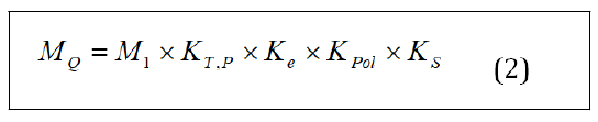
The absorbed dose to water at any reference depth, Zref in water: [2]

For Corrected dosimeter reading at voltage V1
The value of beam quality correcting factor, was corrected for the effect o f t he differences be tween th e re ference beam quality Q0 and the actual user quality Q. The beam Quality Q of a megavoltage photon beam is speci ied with either with tissuephantom ratio or with the Percentage Depth Dose PDD. TPR20,10 is de ined as the ratio of doses on the beam central axis at depths of z = 20 cm and z = 10 cm in water obtained at an SAD of 100 cm and a ield size of 10 cm2 × 10 cm2. TPR20,10 is independent of electron contamination of the incident photon beam.
Results and Discussion
Absolute dosimetry using 1D water phantom measurement
The measurements were conducted using varian clinac iX (manufacture: varian medical system, USA) (varian oncology) at the institute of nuclear medical physics, Bangladesh atomic energy commission [3,4].
It has two photon energies 6 MV and 15 MV and ive electron energies (e.g. 6 MeV, 9 MeV, 12 MeV, 15 MeV and 18 MeV). Absolute dosimetry for 6 MV and 15 MV photon energies were measured by using a 1D water phantom with Farmer Chamber (FC 65-P) for 6MV and 15MV. For this study, 1D phantom, ionization chambers, and electrometers are used.
The ionization chamber is the most practical and most widely used type of dosimeter for accurate measurement of machine output in radiotherapy [5] .
The ionization chamber is put in a water phantom, and is connected with a electrometer. The measurements were done using the Source to Surface Distance (SSD) technique at 10 cm depth with 10 cm2 × 10 cm2 ield size.
Figure 1 shows some experimental set up for the measurement of absorbed to water. Temperature and pressure was found to be T=18.90 C and P=1009 hpa. T0 and P0 from the standards laboratory was T0=200C and P0=1013.25 hpa. The machine was irradiated with a voltage of V=+300 V. For pulsed beam we used the ratio V1/V2=3. We used the calculation for 100 Monitor Unit (MU) with dose rate 400. The chamber calibration factor, ND,W=4.789 Gy/C × 107 Gy/C was taken from chamber calibration certificate. To find out TPR20,10 for 6MV and 15 MV, the ionization chamber was placed at depth of 20 cm with SSD=80 cm, field size 10 cm2 × 10 cm2 and data was taken. Then the ionization chamber was placed at depth of 10 cm with SSD=90 cm, field size 10 cm2 × 10 cm2 and data were taken. Table 1 shows the data for beam quality correction factor TPR20,10 (Table 1).

Figure 1: Experimental set up for absolute dosimetry measurement using 1d water phantom. A) Medical Linac; B) 1D Stand alone water phantom; C) Ionization Chamber: FC 65P; D) Electrometer.
| Energy | Depth=20 cm, SSD=80 cm | Depth=10 cm, SSD=90 cm | ||
|---|---|---|---|---|
| Reading (nC) | Mean (nC) | Reading (nC) | Mean (nC) | |
| 6 MV | 11.42 | 11.415 | 17.1 | 17.1 |
| 11.41 | 17.1 | |||
| 15 MV | 15.27 | 15.275 | 20.13 | 20.13 |
| 15.28 | 20.13 | |||
Table 1: Determination of the value of TPR20,10 for 1D water phantom.
For 6 MV photon beam, TPR20,10 was found 0.667. Using that value KQ was found to be 0.993 from IAEA TRS-398 [2]. For 15 MV photon beam, TPR20,10 was found 0.7588 and KQ was found to be 0.976 To correct the signal some factors are involved. The ionization chamber was set for SSD=100 cm, at 10 cm depth with ield size 10 cm2 × 10 cm2 and data was taken. To correct the signal some factors are involved. Table 2 shows data for determination of M+, M- and M2. M1 was same as M+.
| Energy | M+ (+300 V) | M- (-300 V) | M2 (+100 V) | |||
|---|---|---|---|---|---|---|
| Reading (nC) | Mean (nC) | Reading (nC) | Mean (nC) | Reading (nC) | Mean (nC) | |
| 6MV | 14.3 | 14.3 | 14.3 | 14.305 | 14.17 | 14.17 |
| 14.3 | 14.31 | 14.17 | ||||
| 15MV | 16.76 | 16.76 | 16.77 | 16.77 | 16.5 | 16.505 |
| 16.76 | 16.77 | 16.51 | ||||
Table 2: Determination of M+, M- and M2 for 1D water phantom.
Now the absorbed dose to water for 6 MV photon beam at reference depth, Zref using equation (2) becomes
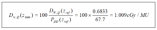
Percentage Depth Dose, PDD for 6 MV photon beam = 67.7, was taken from INMP medical linac data profile which was also similar as other institute [6]. Using this value we can determine Absorbed dose to water for 6 MV photon beam at the depth of dose maximum, Zmax by using [2]
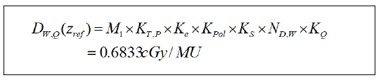
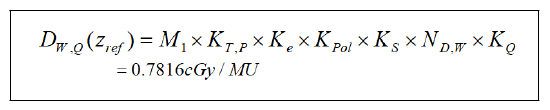
Absorbed dose to water for 15 MV photon beam at reference depth, zref using equation (2)
Percentage Depth Dose, PDD for 15 MV photon beam=76.8, was taken from INMP medical linac data profile which was also similar as other institute [6]. Absorbed dose to water for 15 MV photon beam at the depth of dose maximum, Zmax [2].
Absolute dosimetry using solid water phantom measurement
For the measurement of absorbed to water in case of solid water phantom, temperature and pressure was found to be T=19.50C and P=1000.8 hpa. T0 and P0 from the standards laboratory was T0=200C and P0=1013 hpa. Table 3 shows the data for beam quality correction factor TPR20,10 (Table 3).
| Energy | Depth=20 cm, SSD=80 cm | Depth=10 cm, SSD=90 cmz | ||
|---|---|---|---|---|
| Reading (nC) | Mean (nC) | Reading (nC) | Mean (nC) | |
| 6 MV | 11.07 | 11.075 | 16.77 | 16.77 |
| 11.08 | 16.77 | |||
| 15 MV | 14.81 | 14.815 | 19.67 | 19.67 |
| 14.82 | 19.67 | |||
Table 3: Determination of the value of TPR20,10 for solid water phantom.
For 6 MV photon beam, TPR20,10 was found . Using that value KQ was found to be 0.994 from IAEA TRS-398 [2]. For 15 MV photon beam, TPR20,10 was found and value of KQ was found to be 0.978. Table 4 shows data for determination of M+, M- and M2. M1 was same as M+ (Table 4).
| Energy | M+ (+300V) | M- (-300V) | M2 (+100V) | |||
|---|---|---|---|---|---|---|
| Reading (nC) | Mean (nC) | Reading (nC) | Mean (nC) | Reading (nC) | Mean (nC) | |
| 6 MV | 13.99 | 13.985 | 14 | 14 | 13.88 | 13.88 |
| 13.98 | 14 | 13.88 | ||||
| 15 MV | 16.33 | 16.33 | 16.33 | 16.33 | 16.08 | 16.075 |
| 16.33 | 16.33 | 16.07 | ||||
Table 4: Determination of M+, M- and M2 for solid water phantom.
Now the absorbed dose to water for 6 MV photon beam at reference depth, Zref using equation (2) becomes
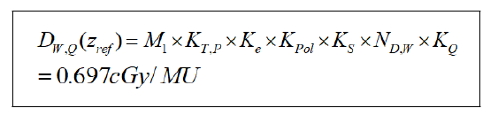
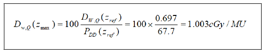
Percentage Depth Dose, PDD for 6 MV photon beam=67.7, was taken from INMP medical linac data profile which was also similar as other institute [6]. Using this value we can determine absorbed dose to water for 6 MV photon beam at the depth of dose maximum, Zmax by using [2].
Absorbed dose to water for 15 MV photon beam at reference depth, Zref using equation (2)
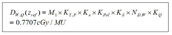
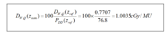
Percentage Depth Dose, PDD for 15 MV photon beam=76.8, was taken from INMP medical linac data profile which was also similar as other institute [6]. Absorbed dose to water for 15 MV photon beam at the depth of dose maximum, Zmax [2].
Assessment the authentication it with machine records
Table 5 shows the comparison of absorbed dose to water at the depth of dose maximum, Zmax for 1D water phantom and solid water phantom and shows the variations it with machine data for 6 MV and 15 MV photon beam.
| Photon energy | Machine data | 1D water phantom | Deviation | Solid water phantom | Deviation |
|---|---|---|---|---|---|
| 6MV | 1cGy/MU | 1.009 cGy/MU | 0.90% | 1.003 cGy/MU | 0.30% |
| 15MV | 1cGy/MU | 1.0177 cGy/MU | 1.77% | 1.0035 cGy/MU | 0.35% |
Table 5: Absorbed dose (at the depth of dose maximum, Zmax) variation of 1 D water phantom and solid water phantom with the machine data.
Conclusion
Absolute dosimetry for photon energies was determined using the IAEA Technical Report Series (TRS-398). The machine was tuned to deliver 1cGy/MU at SSD technique at Dmax for all the available photon energies and kept as a baseline. Beam quality index were measured for all the energies using the TPR20,10 and absolute dose determination were done at 10 cm depth using SSD technique. All the measurements were carried out using the IBA 1D water phantom and solid water phantom with farmer 0.65 cc (FC 65-P) chamber. The meter readings were corrected for temperature-pressure correction, polarity effect and recombination effect. The corrected meter readings were multiplied using the absolute dose to water calibration factor (ND,W,Q0) provided by the secondary standard dosimetry lab along with the appropriate KQ according to the beam quality. Absorbed dose to water at reference depth (10 cm) for 1D water phantom for 6 MV and 15 MV photon beam was found to be 0.68 cGy/MU and 0.78 cGy/MU. Absorbed dose to water at the depth of dose maximum (zmax) for 6 MV and 15 MV Photon Beam was found to be 1.009 cGy/MU and 1.0177 cGy/MU. For solid water phantom absorbed dose to water at reference depth (10 cm) for 6 MV and 15 MV photon beam was found to be 0.697 cGy/MU and 0.77 cGy/MU. Absorbed dose to water at the depth of dose maximum (zmax) for 6 MV and 15 MV photon beam was found to be 1.003 cGy/MU and 1.0035 cGy/MU.
For 1 D water phantom, a variation with machine data was obtained as 0.9% and 1.77% for 6 MV and 15 MV photon beam respectively. For Solid Water phantom, a variation with machine data was obtained as 0.3% and 0.35% for 6 MV and 15 MV photon beam respectively. The beam quality parameters of the available photon beams were found to be Varian acceptance tolerance and also compatible with the IAEA TRS 398 recommendations.
Acknowledgement
The authors would like to thank ministry of science and technology, Government of Bangladesh for providing the ADP project entitled as “Establishment of Institute of Nuclear Medical Physics”. The authors also would like to thank all the personnel of the Institute of Nuclear Medical Physics (INMP), Bangladesh atomic energy commission for allowing us to use the VARIAN Clinac iX and give necessary support for this work.
References
- Shalek RJ (1977) Determination of absorbed dose in a patient irradiated by beams of × or gamma rays in radiotherapy procedures. Medical Physics.
[Crossref], [Google Scholar]
- Stephen VM (2001) Absorbed dose determination in external beam radiotherapy: an international code of practice for dosimetry based on standards of absorbed dose to water. IAEA TRS-398. Health Physics 81: 592-593.
- Roy SK, Das PK, Khatun R, Rahman MA, Akter S, et al. (2021) Dosimetric characteristics of 6 MV medical linac at BAEC. IJMPCERO 10: 38-46.
[Crossref], [Google Scholar]
- Khatun R, Rana M, Ahasan M, Akter S, Uddin F, et al. (2017) Site planning of a newly installed LINAC at BAEC, Bangladesh. Univers J Med Sci 5: 8-12.
- Determination of absorbed dose in a patient irradiated by beams of × or gamma rays in radiotherapy procedures
- Mani KR, Bhuiyan MA, Rahman MS, Islam MSA (2018) Open beam dosimetric characteristics of true beam medical linear accelerator With Flattening Filter (WFF) and Flattening Filter Free (FFF) beam. Polish J Medical Phys Engineering 24:79-89.
[Crossref], [Google Scholar]

Open Access Journals
- Aquaculture & Veterinary Science
- Chemistry & Chemical Sciences
- Clinical Sciences
- Engineering
- General Science
- Genetics & Molecular Biology
- Health Care & Nursing
- Immunology & Microbiology
- Materials Science
- Mathematics & Physics
- Medical Sciences
- Neurology & Psychiatry
- Oncology & Cancer Science
- Pharmaceutical Sciences