Evaluation of Radiation Dose Received by Paediatric Patients during Routine XRay Examinations of the Chest in Three Selected Hospitals in Cameroon
DOI10.36648/2574-285x.7.4.015
Chi Emmanuel*, O N Samba, C Tetchoka Manemo and L C Fai
Department of Physics, University of Dschang, Dschang, Cameroon
- *Corresponding Author:
- Chi Emmanuel
Department of Physics,
University of Dschang, Dschang,
Cameroon,
Tel: 237 676417756;
Email: chiemmanuel5@gmail.com
Received: 13-April-2022, Manuscript No. M-3793; Editor assigned: 16-April-2022, PreQC No. P-3793; Reviewed: 30-April-2022, QC No. Q-3793; Revised: 31-May-2022, Manuscript No. R-3793; QI No. Q-3793 Published: 28-June-2022 DOI: 10.36648/2574-285x.7.4.015
Citation: Emmanuel C, Samba ON, Manemo CT, and Fai LC (2022) Evaluation of Radiation Dose Received By Paediatric Patients during Routine XRay Examinations of the Chest in Three Selected Hospitals in Cameroon. J Med Phys Appl Sci Vol: 7 No: 4:15.
Abstract
The radiation risks associated with children are higher than the risks for adults. Children have growing organs and they have a longer life expectancy than that of adults. Consequently, the effects of damage from radiation could be greater in children than in adults. This study sought to measure the mean Entrance Surface Dose (ESD) and third quartile values for paediatric chest X-ray examinations, compare these to findings and recommendations from other studies and propose methods of dose reduction in three hospitals (H1-H3) in Cameroon. The age groups considered in this study were <1 year, 1-<5 years, 5-<10 years and 10-15 years. The mean ESD for the chest AP in the age range <1 year in the three hospitals (H1-H3) were respectively 0.10, 0.09, 0.66 mGy; for the age 1-<5 years were respectively 0.12, 0.10, 0.48 mGy and for the age range 5-<10 years were respectively 0.13, 0.09, 0.34 mGy. According to NRPB 2000, the reference levels are given in defined age that is (newborn, 1 year, 5 years, and 10 years). For example, for AP and PA chest radiographs; 0.05 mGy for newborns and 1 year old, 0.07 mGy for 5 years and 0.12 mGy for 10 years old children. It is difficult to compare this reference with the results of this study where age range had been used to evaluate the mean ESD values but can still provide useful information for comparison. A weak correlation was also found between ESD and weight/age. The fluctuation of dose from one hospital to another is an indication that there is a need for optimization in paediatric radiography. This can be done through Quality Assurance Programs (QAP).
Keywords
Entrance surface dose; Paediatric; Optimization
Introduction
Living things have evolved in an environment that has significant levels of ionizing radiations. Radiation is used in medical diagnosis and treatment of diseases such as cancer. Thus, there are a lot of benefits from the careful use of radiations (X-rays). Despite these benefits, the use of medical Xrays still have hidden problems [1]. The prevention of the hazardous effects of ionizing radiation has been a critical concern despite their valuable contribution in medical imaging to diagnosis and subsequent treatment of various diseases [4]. Radiation protection in paeditric radiology requires special attention than in adult radiology because children are more sensitive to radiation and at higher risk than adults [14]. The higher risk in children is explained by their longer life expectancy, which allows more time for any harmful effects of radiation to manifest; and the fact that developing organs and tissues are more sensitive to the effects of radiation [2]. The importance of constant monitoring of the radiation dose received by paediatrics during a radiological examination cannot be understated as studies done indicate that risk for radiation related cancer in children is a result of early life exposure to ionizing radiation. On this note, Quality Assurance Programs (QAP) should be implemented in Paeditric radiology. Quality assurance in paediatric radiology is more important, since it is known that children are more sensitive to radiation than adults. Therefore, in this case, closer attention should be paid to improve the diagnostic information, reducing the child dose as much as possible (K. Schneiden, 1995). The use of Diagnostic Reference Levels (DRLs) is a very useful and applicable method in many countries as a measure to reduce over exposure or under exposure. DRLs are set based on the survey of different hospitals using different X-ray tubes or machines [3,4].
From the findings of the researchers’, there are no radiation dose survey measures, for both adults and paeditrics at the studied Hospitals and so therefore there is a possibility of over exposure. The National Agency for Radiation Protection (NARP) in Cameroon which is responsible for radiation protection of patients, general public and staff have not yet set baseline dose which are applicable to each hospital. A dosimetric survey was therefore important to ensure that children are not put at higher risk of developing radiation-induced cancer [5]. The purpose of this article is to report on a study that sought to investigate the radiation dose received by children (1 to 15 years) in terms of ESD, evaluated using mathematical models from the data obtained from the patients and the exposure parameter from the X-ray machine during Exposure for paeditric chest, in three hospitals in Cameroon. From the results we shall establish a correlation between ESD, weight, height and age and comparing them with those reported in literature [6].
Material and Methods
This study was carried out to estimate, using mathematical models, the ESD received by paeditric patients referred to diagnostic chest X-rays in three hospitals in Cameroon [7]. These three hospitals were chosen because they receive a large number of patients. A total of 232 radiographs were obtained from children within the age of 1 week to 15 years, 115 females and 117 males. A total of three X-ray machines were used in this study with characteristics summarized in Table 1.
| Hospital | Equipment model | Date of manu-facture | Date of installation | Serial number | Film screen type/speed | Total filtration |
|---|---|---|---|---|---|---|
| H1 | GE Healthcare | May-2011 | 2012 | 75422HL6 | Fujifilm/400 | 2.5 mmAl |
| H2 | Stepanix | May-2011 | 2012 | 11H411 | Fujifilm/400 | 3.09 mmAl |
| H3 | CGR/ATAK25 | 1968 | 1974 | 6IR659 | Kodac/200 | 2.93 mmAl |
Table 1: Characteristics of the investigated X-rays machines.
The measurement of the tube output was performed using a calibrated DIAVOLT universal all in one Quality Control (QC) meter. It is a non-invasive X-ray meter for kV, dose and irradiation time measurements at X-ray installations. It was used in the absence of the patients in this study to obtain the tube output of the equipment [8]. Two examination techniques were taken in account: PA and AP. Radiological parameters such as kilovoltage (kV), product of X-ray tube current and time (mAs) and Focus-to-Skin Distance (FSD) with the patient characteristics such as weight, height and age were recorded with the assistance of the technician or engineer on duty. The type of Xray examination and the view (projection) were obtained from the patient request form. The patient’s age was asked from patient care taker, the height was measured using a measuring tape and the weight of the patient was obtained using a balance. Information such as weight and age were also verified from their medical booklets. During the examination, the kV, and mAs were directly read from the control console and the tape which was fixed on the X-ray tube helped us to measure FSD directly after the patient was positioned for the examination. The ESD received by paediatric patients was evaluated using equation 1 with a backscatter factor of 1.35.
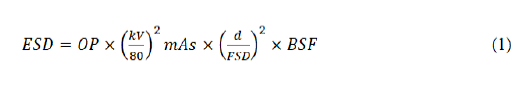
Where
BSF is the backscatter factor
OP is the output in mGy/mAs of the X-ray tube at 80 kV at distance of 100 cm normalized to 20 mAs
mAs is the time current product measured in mAs and
FSD is the focus to skin distance
The data collected was entered and analyzed using excel. The results of the min, max, mean, 3rd quartile and standard deviation are presented in Table 3.
Results
The male patients were 50.43% (n=117) of the total population while the female where 49.57% (n=115) of the total population. The patients were divided into four groups separate age groups: <1 yr, 1-<5 yrs, 5-<10 yrs and 10-15 yrs. This was done due to the wide variation in body sizes common with paediatric patients. The separation of the patients into different age groups made it easy for the calculation of the ESD [10]. Patient demographic data and exposure parameters for this study are summarized in Table 2, while the results of the ESD obtained from this study for the chest PA and AP examinations are displayed in Table 3.
| Weight/kg | Height/cm | kV | mAs | Age | |
|---|---|---|---|---|---|
| Minimum | 3.5 | 27 | 40 | 0.97 | 1 day |
| Mean | 15.75 | 163 | 75.51 | 6.59 | 2.56 |
| Maximum | 62 | 74.86 | 120 | 30 | 15 |
| Standard deviation | 11.11 | 32.85 | 26.85 | 7.18 | 3.64 |
Table 2: Summary of patient demographic and exposure parameters used in this study.
| Projection | Studied centres | ESD (mGy) | |||||
|---|---|---|---|---|---|---|---|
| Mean | Min | Max | Median | 3rdqtle | SD | ||
| <1 years | |||||||
| AP | H1 | 0.1 | 0.07 | 0.34 | 0.07 | 0.07 | 0.08 |
| H2 | 0.09 | 0.03 | 0.2 | 0.09 | 0.1 | 0.04 | |
| H3 | 0.66 | 0.35 | 1.06 | 0.67 | 0.75 | 0.17 | |
| 1-<5 years | |||||||
| AP | H1 | 0.12 | 0.07 | 0.35 | 0.1 | 0.13 | 0.08 |
| H2 | 0.1 | 0.06 | 0.14 | 0.11 | 0.12 | 0.04 | |
| H3 | 0.48 | 0.19 | 0.86 | 0.49 | 0.57 | 0.23 | |
| PA | H1 | 0.09 | 0.07 | 0.13 | 0.07 | 0.11 | 0.03 |
| H2 | 0.1 | 0.06 | 0.17 | 0.09 | 0.12 | 0.04 | |
| H3 | 0.32 | 0.18 | 0.49 | 0.32 | 0.35 | 0.08 | |
| 5-<10 years | |||||||
| AP | H1 | 0.13 | 0.11 | 0.14 | 0.14 | 0.14 | 0.02 |
| H2 | 0.09 | 0.06 | 0.16 | 0.08 | 0.08 | 0.04 | |
| H3 | 0.34 | 0.3 | 0.38 | 0.32 | 0.36 | 0.03 | |
| PA | H1 | 0.13 | 0.1 | 0.19 | 0.13 | 0.15 | 0.03 |
| H2 | 0.08 | 0.03 | 0.14 | 0.07 | 0.08 | 0.04 | |
| H3 | 0.35 | 0.19 | 0.48 | 0.36 | 0.37 | 0.06 | |
| 10-15 years | |||||||
| PA | H1 | 0.2 | 0.14 | 0.26 | 0.19 | 0.22 | 0.03 |
| H2 | 0.12 | 0.04 | 0.2 | 0.15 | 0.17 | 0.06 | |
| H3 | 0.5 | 0.3 | 1.59 | 0.44 | 0.52 | 0.27 | |
| AP | H2 | 0.13 | 0.11 | 0.14 | 0.14 | 0.14 | 0.02 |
Table 3: Statistical distribution of ESD (in mGy) for the three hospitals and age ranges.
The ESD for chest AP was plotted against weight, age, mAs and kV and the scatter diagrams of Figures 1 to 4 were obtained.

Figure 1: ESD plotted against weight.

Figure 2: ESD plotted against age.

Figure 3: ESD plotted against mAs.

Figure 4: ESD plotted against kV.
It shows that there was no significant correlation between ESD and weight (r=-0.13292), between ESD and age (r=-0.2249) and between ESD and kV (r=-0.71256). However, Figure 3 shows a significantly strong correlation between ESD and mAs (r=0.84564).
Correlation between ESD and weight (r=-0.132) between ESD and age it was (r=-0.2249). Thus there was no signi icant correlation between ESD and weight/age.
Discussion
The ESD for of 232 patients were evaluated and the results presented in Table 3. A mean dose of 0.12 μGy was obtained for the chest AP for children in the age group <1 year. This value is slightly higher compared to a recommended value of 0.05 μGy by the NRPB. The mean values of the ESD obtained from H1 and H2 for the age group 1-<5 years compare favourably with the recommended values by the NRPB and EC, while those from H3 were higher than the recommended values (Table 4 and Figure 5) and those from similar literature [9]. The luctuation in dose could be due to factors such as state of equipment and the technique or protocol used. The radiation dose received by patients is greatly in luenced by the equipment used and the protocol applied [10]. It can be seen from Table 1 that the X-ray machine of H3 is about 50 years old from the year of manufacture to the time of this study, meaning according to the suspension levels for diagnostic radiography, the tube of H3 should be suspended. Besides, the ilm speed is extremely low; meaning high doses of radiations will be delivered to patients to have a good image quality. Thus, from all indications, patients will receive high doses than normal from H3 [11].
| Projection | Current study | National Radiological Protection Board | EC | Nigeria, 2013 |
|---|---|---|---|---|
| <1 year | ||||
| Chest AP | 0.28 | 0.05 | ||
| 1-5 years | ||||
| Chest AP | 0.23 | 0.07 | 0.1 | |
| Chest PA | 0.17 | 0.07 | 0.1 | 0.11 |
| 5-<10 years | ||||
| Chest AP | 0.19 | 0.12 | ||
| Chest PA | 0.19 | 0.12 | 0.159 | |
| 10-15 years | ||||
| Chest AP | 0.13 | |||
| Chest PA | 0.27 | 0.62 | ||
Table 4: Comparison of the mean ESD obtained from this study with reference.

Figure 5: Comparison of the meanof ESD from the three hospitals with reference values of NRPB and EC.
The quantity of radiation dose received by a patient is greatly affected by the radiographic technique used.
Each hospital has its own protocol for every examination, for example at H3 theFSD was in the range 70-87 cm, meanwhile at H1 and H2 the FSD were respectively in the ranges 74-133 cm and 62-132 for the age group <1 year [12]. This difference in FSD has an effect on patient dose and the doses reported by different authors. Low kV and high mAs technique are also attributing factors to dose received by a patient. A study carried out in Nigerian hospitals showed that those centers which used this technique obtained higher ESD values compared to those that used high kV and low mAs. This is the other reason why ESD values were high at H3 as low kV (42-75 kV) and high mAs (5-30 mAs) values were used throughout the study [13,14].
We expected from this study that as the weight/age of the patients increase, the ESD should also increase. This was not the case from our indings. There was a large luctuation in ESD with weight/age (Figures 1 and 2). The correction coefficient between ESD and weight was (r=-0.13292) and between ESD and age it was (r=-0.2249). Thus there was no signi icant correlation between ESD and weight/age [15]. The luctuation in dose weight/age could to due variation in patient size which is common with paeditric patients. Besides, there was no signi icant relationship between ESD and kV as the correlation coefficient between kV and ESD was found to be (r=-0.71256). However, there was a signi icantly strong correlation between mAs and ESD (r=0.84564), as expected.
Conclusion
Diagnostic radiology is the greatest source of public exposure to radiation. Although are medically justi iable, it is important to optimise the dose per examination (BASSON, J.K et al.) especially when dealing with paediatrics. Due to the cell sensitivity of children to ionizing radiation, special attention is required in radiation protection of paediatrics patients than in adults. The United Nation Scienti ic Committee on Effects of Atomic Radiation (UNSCEAR) reported that children exposed to radiation at the early age of less than 5 years are twice to thrice sensitive to effects of radiation than adults. These results suggest that pediatric patients especially below the age of 5years may be receiving higher radiation dose compared to the recommended doses. This could be due to the lack of adequate quality control measures on the equipment and the age of the machines and radiographic techniques applied.
Recommendations
Results obtained in this study may act as a baseline for the establishment of local DRLs for chest examinations and they serve as a guideline in improving radiographic practice. However, due to the limitations in this study, the researchers recommend further studies in different hospitals as that may yield a more reliable mean ESD and third quartile value. The researchers also recommend careful implementation of the ALARA principle, application of more appropriate radiographic technique which is relevant to paediatric patients, the setting up centres dedicated for paeditric examinations and to create awareness on suspension levels in diagnostic radiology.
References
- Ademola AK, Obed RI, Adejumobi CA, Abodunrin OP, Alabi OF, et al. (2013) Assessment of entrance skin dose in routine x-ray examinations of chest, skull, abdomen and pelvis of children in five selected hospitals in Nigeria. IOSR J Appl Phys 5: 47-50.
- ARPANSA R (2008) Code of practice for radiation protection in the medical applications of ionizing radiation. Yallambie: Australian Radiation Protect Nuclear Safety Agency.
- Basson JK, Swiegers WRS (1999) SA Forum for radiation protection. Occupat Health 5: 40-46.
- Kim BH, Do KH, Goo HW, Yang DH, Oh SY, et al. (2012) National survey of radiation doses of pediatric chest radiography in Korea: Analysis of the factors affecting radiation doses. Korean J Radiol 13: 610-617.
[Crossref] [Google Scholar] [PubMed]
- Egbe NO, Inyang SO, Ibeagwa OB, Chiaghanam NO (2008) Pediatric radiography entrance doses for some routine procedures in three hospitals within eastern Nigeria. J Med Phys 33: 29.
[Crossref] [Google Scholar] [PubMed]
- Simeonov G (2013) Criteria for acceptability of medical radiological equipment in Euratom legislation. Radiat Prot Dosimetry 153: 147-149.
[Crossref] [Google Scholar] [PubMed]
- Fendel H, Schneider K, Schofer H, Bakowski C, Kohn MM (1985) Optimisation in paediatric radiology. Are there specific problems for quality assurance in paediatric radiology. In Criteria and methods for quality assurance in medical X-ray diagnosis. Proceedings of the scientific seminar held in Udine, Italy, 17-19 April 1984.
- International Commission on Radiation Protection, ICRP (2011) Radiological protection in paeditric diagnostic and interventional radiology. Annal ICRP 4839: 3982-4649.
- Schneider K (1995) Evolution of quality assurance in paediatric radiology. Radiat Protect Dosimetry 57: 119-123.
- Kiljunen T, Jarvinen H, Savolainen S (2007) Diagnostic reference levels for thorax X-ray examinations of paediatric patients. Br J Radiol 80: 452-459.
[Crossref] [Google Scholar] [PubMed]
- Kleinerman RA (2006) Cancer risks following diagnostic and therapeutic radiation exposure in children. Pediatr Radiol 36: 121-125.
[Crossref] [Google Scholar] [PubMed]
- Mohamadain KEM, Azevedo ACP (2009) Radiation dose survey in conventional pediatric radiology. J Sci Tech 10: 175-183.
- Olowookere CJ, Babalola IA, Olayiwola MO, Odina G, Obed RI, et al. (2009) Comparison of five models for assessing patient dose from radiological examinations. Afr J Med Phys Biomed Eng Sci 1: 21-29.
- Suliman II, Elshiekh EHA (2008) Radiation doses from some common paediatric X-ray examinations in Sudan. Radiat Prot Dosimetry 132: 64-72.
[Crossref] [Google Scholar] [PubMed]
- United Nations (2000) Sources and effects of ionizing radiation, Report to the General Assembly with scientific annexes.

Open Access Journals
- Aquaculture & Veterinary Science
- Chemistry & Chemical Sciences
- Clinical Sciences
- Engineering
- General Science
- Genetics & Molecular Biology
- Health Care & Nursing
- Immunology & Microbiology
- Materials Science
- Mathematics & Physics
- Medical Sciences
- Neurology & Psychiatry
- Oncology & Cancer Science
- Pharmaceutical Sciences