Comparison of Absorbed Dose Rate in the Healthy Organs by the Mammosite and Contura Applicators in Breast Brachytherapy
Fatemeh Mahdavi*, Alireza Shirazi Hosseini Dokht and Mahdi Sadeghi
Published Date: 2025-02-28Fatemeh Mahdavi1*, Alireza Shirazi Hosseini Dokht2 and Mahdi Sadeghi3
1Department of Medicine, Tehran University of Medical Sciences, Tehran, Iran
2Department of Biophysics, University of Tehran, Tehran, Iran
3Department of Medical Physics, School of Medicine, Iran University of Medicine Science, Tehran, Iran
*Corresponding Author:
- Fatemeh Mahdavi
Department of Medicine, Tehran University of Medical Sciences, Tehran, Iran
E-mail:fatemahdavi@yahoo.com
Received date: October 07, 2023, Manuscript No. IPIMP-23-17984; Editor assigned date: October 09, 2023, PreQC No. IPIMP-23-17984 (PQ); Reviewed date: October 23, 2023, QC No. IPIMP-23-17984; Revised date: January 09, 2025, Manuscript No. IPIMP-23-17984 (R); Published date: January 16, 2025, DOI: 10.36648/2574-285X.10.1.90
Citation: Mahdavi F, Dokht ASH, Sadeghi M (2025) Comparison of Absorbed Dose Rate in the Healthy Organs by the Mammosite and Contura Applicators in Breast Brachytherapy. J Med Phys Appl Sci Vol:10 No:1
Abstract
Brachytherapy is one of the treatments for breast cancer. In brachytherapy treatment systems, high dose rate sources are used in interstitial implantation. By the late 1960’s, with a few small exceptions, all brachytherapy sources and applicators were manually connected or implanted inside the patient.
With increasing concerns about the quality of treatment and the possible effects of radiation on employees, many efforts have been made to reduce radiation exposure to operators by using after loading machines. The after loading method requires the initial placement of non-radioactive applicators or carriers inside or on the patient's body, after which radioactive materials are placed. In this method, the sources move with a specific radioactivity at certain distance and precisely to each other in the systems after loading (after load).
Iridium 192 was first used for Brachytherapy at the University of Oxford in 1976 and has been widely used in this field. Accurate knowledge of dose distribution is of particular importance to design treatment methods.
In this study, modeling of two standard applicators in the breast cancer treatment named Mammosite and Mammosite-ML (Contura) is done by Monte Carlo simulation code.
Then, the amount and distribution of the dose in breast brachytherapy the dose reached to the organs at risk of radiation, including; lungs, ribs, and skin and other organs, are evaluated, and calculated and the time of treatment in each session is checked to introduce the best applicator with the lowest dose in healthy organs and the shortest duration of treatment.
Keywords
Brachytherapy; Mammosite; Contura; Breast; Monte Carlo simulation
Introduction
Treatment for breast cancer varies according to the cancer classification, especially considering the size of the tumor, the extent of the tumor spread, and the risk of recurrence. Published protocols that suggest specific treatments for breast cancer that fall into a particular category. Treatments include; surgery, systematic therapies (chemotherapy and hormone therapy), and radiation therapy, its types. Brachytherapy is one of the ways to treat breast cancer. Brachytherapy is a short-term treatment for cancer with radiation from encapsulated small radionuclide sources. This type of therapy is performed by direct placement of sources within or near the treated volume; then the dose is continuously delivered to the treatment site. In this study, the seeds of iridium-192, which is the isotope iridium-191, was used [1].
Iridium-192 is produced by the neutron activity of iridium-191, and has a half-life of 74 days. Photon radiation is a complex spectrum with an average energy of 0.38 MeV. In the past, radioactive sources were usually manually loaded into applicators or catheters targeted within the target volume. At the end of treatment, the sources were removed again manually. These processes expose employees. Today, computercontrolled remote after load systems have been developed to minimize this exposure.
In this research, the aim is to simulate Mammosite and Contura applicators using Monte Carlo simulation code. Using the female model ORNL-MIRD (Oak Ridge National Laboratory- Medical Internal Radiation Dose) phantom, a hypothetical tumor or target volume is placed in the breast tissue at a distance of 1 cm from the applicator balloon and then the amount of absorbed dose in the lungs, ribs and skin and other organs is determined.
The purpose of this study is to evaluate the amount of absorbed dose in OAR and other organs and to determine the duration of treatment in each session by each applicator and to determine the applicator with the ability to generate asymmetric dose [2].
Materials and Methods
The MCNPX code is based on the Monte Carlo method. The MCNP code is a robust computational multi-purpose code used to transport neutrons, electrons, and photons. MCNP can also calculate quantities in complex and different geometries. In this study, the simulation was performed using MCNPX code version 2.6.
The ORNL-MIRD Phantom is an analytical model of the human body by ORNL-MIRD Publications. In this phantom, all the human body organs with different geometries are represented by analytical equations. The MIRD phantom provides an analytical model of the human body. Calculating the dose in the organs requires a detailed description of the organs, geometry and the chemical structure of the tissue.
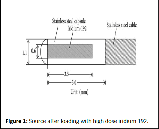
The source used in this research is 192 iridium springs. This source is in the form of a platinum capsule, its inner and outer diameters are 0.0325 cm and 0.045 cm, respectively. This source emits beta radiation with 672 keV energy and gamma rays with different energies, approximately 468 keV. The activity of the source used is equal to 10 Ci, and its specific activity is about 450 Ci/g, and the average energy is 380 keV. Its half-life (73.8 days) is about 74 days. The source simulated here is a cylinder with a length of 0.5 and a radius of 0.017 cm (Figure 1).
Figure 1: Source after loading with high dose iridium 192.
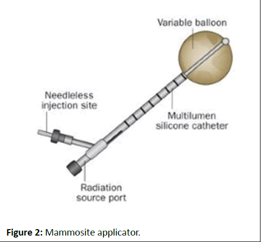
Mammoth single duct balloon, this device has a catheter connected to an HDR afterload machine to connect to the source [3]. The end of the catheter is surrounded by a balloon 4 to 5 cm in diameter that can be filled with saline solution or other material. The balloon is placed in the lumpectomy cavity of the patient's body, which can give a dose of 34 Gy to the tissue from the applicator, surface. The advantage of this device is that easy to use (Figure 2) [4,5].
Figure 2: Mammosite applicator.
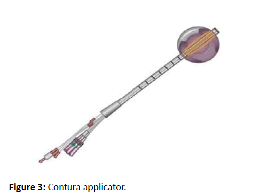
Other multichannel tools, such as the Mammosite-ML, are almost identical to the Mammosite; the difference is that Mammosite has a central main duct [6-9]. Still, Mammosite-ML has three additional ducts around the central duct, which are 120 degrees apart. Ducts added to Mammosite-ML give the ability to shape the dose. Another model of Mammosite-ML is the Contura applicator, which is one type has four ducts around the central duct at a distance of 90 degrees from each other. The ducts have a higher degree of curvature than the previous model, and in the best model it has ive ducts that are used to enter the radioactive source to cover more space, in this study, this model is used for simulation. The maximum distance between the central duct and other ducts is 5 mm. Like Mammosite is available in, 4 sizes 4-5 cm and 5.4-6 cm, these dimensions enable the Contura to have the same volume as the Mammosite applicator (Figure 3).
Figure 3: Contura applicator.
Results
Using Monte Carlo simulation, two applications, Mammosite and Contura, are simulated. Both applicators were placed in the right and left breasts, and a target volume or hypothetical tumor was placed 1 cm from the applicators to calculate the absorbed dose by both the target volume and all organs. The time required to reach the prescribed dose in each treatment fraction is also calculated, and then the dose in each treatment fraction is calculated with the target volume and the treated breast as well as the lungs, ribs, and skin organs, which is the main goal of this study [10].
In this study, the F6 Tally was used to calculating the dose of different organs. Which estimates the deposited energy. (F6 (MeV/g/particle source)). In this study, calculations were performed for 108 particles per date to reduce the error rate.
Mammosite applicator simulation results by placing the target volume on the right and left breasts:
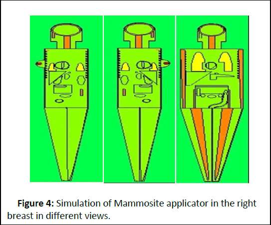
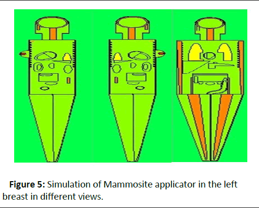
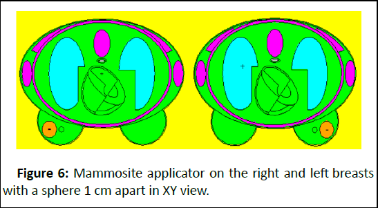
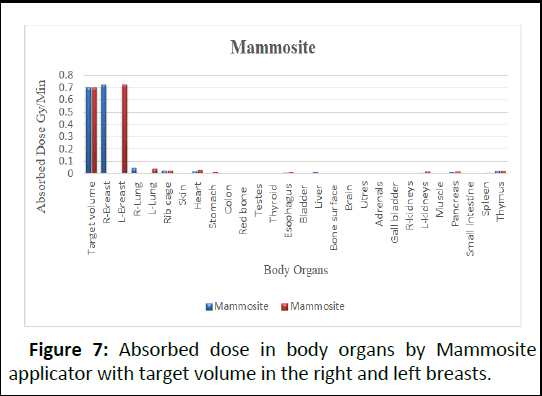
The following results are obtained by considering the Mammosite applicator and the source of iridium 192, and the target volume in the patient's right and left breasts. The amount of absorbed dose in the target organs has been calculated. The following Figures 4-7 shows the simulated geometry.
Figure 4: Simulation of Mammosite applicator in the right breast in different views.
Figure 5: Simulation of Mammosite applicator in the left breast in different views.
Figure 6: Mammosite applicator on the right and left breasts with a sphere 1 cm apart in XY view.
Figure 7: Absorbed dose in body organs by Mammosite applicator with target volume in the right and left breasts.
As expected, the absorbed dose is the highest in the target volume or hypothetical tumor, and the treated breast, and then the lungs have a significant dose.
Given that the prescription dose in High Dose Rate brachytherapy (HDR) is 34 Gy per 10 fractions of treatment that is in each treatment fraction, a dose of 3.4 Gy should be absorbed into the target volume [11]. The time required to reach a dose of 3.4 Gy in each treatment fraction is calculated as follows:
The absorbed dose in the target volume by MCNP=0.7 Gy/min
The time required to reach the base dose=3.4 Gy ÷ 0 .7 Gy/min=4.85 ≈ 5 min
Multiplying the results of the absorbed dose by 5 minutes, the absorbed dose of the organs is obtained by the Mammosite applicator in one treatment fraction. The critical organs in this research are identified in the Table 1 below.
| Body organs | Target volume in right breast | Target volume in left breast |
|---|---|---|
| Target volume | 3.505 | 3.51 |
| Right breast | 3.63 | 0.0344 |
| Left breast | 0.03445 | 3.63 |
| Right lung | 0.229 | 0.0302 |
| Left lung | 0.0287 | 0.22 |
| Ribs | 0.13 | 0.13 |
| Skin | 0.04675 | 0.04675 |
Table 1: Absorbed dose (Gy) of body organs in a treatment fraction with Mammosite applicator.
Contura applicator simulation results by placing the target volume on the right and left breasts:
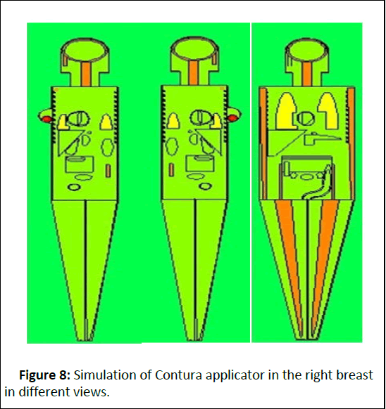
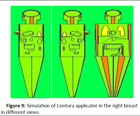
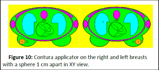
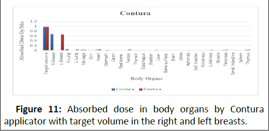
The following results are obtained by considering the Contura applicator and the source of iridium 192, and the target volume in the patient's right and left breasts. The amount of absorbed dose in the target organs has been calculated. The following Figures 8-11 shows the simulated geometry [12].
Figure 8: Simulation of Contura applicator in the right breast in different views.
Figure 9: Simulation of Contura applicator in the right breast in different views.
Figure 10: Contura applicator on the right and left breasts with a sphere 1 cm apart in XY view.
Figure 11: Absorbed dose in body organs by Contura applicator with target volume in the right and left breasts.
The time required to reach a dose of 3.4 Gy per fraction, as in the previous section, is calculated as follows:
The absorbed dose in the target volume by MCNP=0.97 Gy/min
The time required to reach the base dose=3.4 Gy ÷ 0.97 Gy/ min=3.48 ≈ 3.5 min
Multiplying the results of the absorbed dose by 5 minutes, the absorbed dose of the organs is obtained by the Contura applicator in one treatment fraction. The critical organs in this research are identified in the Table 2 below.
| Body organs | Target volume in right breast | Target volume in left breast |
|---|---|---|
| Target volume | 3.41 | 3.4 |
| Right breast | 2.38 | 0.0371 |
| Left breast | 0.0364 | 2.31 |
| Right lung | 0.185 | 0.0269 |
| Left lung | 0.0252 | 0.1568 |
| Ribs | 0.11 | 0.11 |
| Skin | 0.035 | 0.035 |
Table 2: Absorbed dose (Gy) of body organs in a treatment fraction with Contura applicator.
According to the results of Table 3 in the Mammosite applicator, the ratio of the absorbed dose of the treated breast tissue to the absorbed dose by the target volume for the right and left breasts is 103%. Therefore, it can be concluded that the same dose that reached the tumor tissue hit the healthy tissue of the treated breast to the same extent, and this indicates a symmetrical dose and proves that the Mammosite applicator has a symmetrical dose distribution.
| Applicator | Right breast | Left breast | Right lung | Left lung | Ribs | Skin |
|---|---|---|---|---|---|---|
| Mammosite Right breast |
103.50% | 0.90% | 6.50% | 0.80% | 3.70% | 1.30% |
| Mammosite Left breast |
0.90% | 103.30% | 0.80% | 6.20% | 3.70% | 1.30% |
| Contura Right breast |
69% | 1.06% | 5.40% | 0.70% | 3.20% | 1.02% |
| Contura Left breast |
1.08% | 67% | 0.70% | 4.50% | 3.20% | 1.04% |
Table 3: Results from the percentage of absorbed dose ratio in organ at risk to the target volume.
But in the Contura applicator, this ratio is 69% and 67% for the right and left breasts, respectively which indicates a lower ratio. Therefore, it can be concluded that the same dose that reached the tumor tissue did not reach the same dose to the healthy tissue of the treated breast, and this indicates an asymmetric dose and proves that the Contura applicator is capable of asymmetric distribution of the dose.
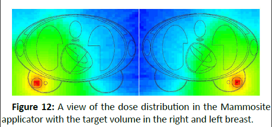
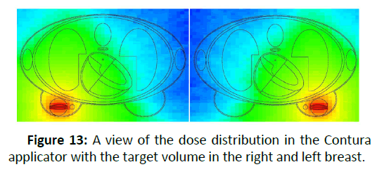
According to the results of the table above, it was found that the lower the ratio of absorbed dose in healthy organs to the target volume, the better and that applicator will be more capable and can provide safer and better treatment. Figures 12 and 13 show a dose distribution in Mammosite and Contura applicators.
Figure 12: A view of the dose distribution in the Mammosite applicator with the target volume in the right and left breast.
Figure 13: A view of the dose distribution in the Contura applicator with the target volume in the right and left breast.
In Figures 12 and 13, it is evident that the red areas have the highest dose, which includes the applicator itself, the target volume, and the treated breast. As the distance increases, the intensity of the dose decreases. There is still a significant, dose in the yellow areas and the green areas have absorbed a higher dose than the blue areas.
Discussion
According to the simulation results, the Mammosite applicator is incapable of asymmetric dose distribution, and healthy organs around the breast, including the lungs, ribs, and skin, receive significant doses. Still, the Contura applicator can cause asymmetric dose distribution and create a safer and higher quality treatment. Therefore, the research hypothesis that increasing the number of ducts, the ability to produce asymmetric doses increases, and the treatment will be better is correct.
The Monte Carlo code can estimate the error rate. In this study, the error rate in determining the absorbed dose for all organs in both applicators for the right and left breast is less than 5%, which indicates the appropriate and acceptable accuracy of the results.
Conclusion
According to the calculations, the time required to reach the dose of 3.4 Gy in each treatment session with the Mammosite applicator is about 5 minutes. With the Contura applicator, it is about 3.5 minutes, which shows the superiority of the Contura applicator over the Mammosite applicator because, in a shorter time, the same dose prescribed in each treatment session can reach the target volume. Of course, the Mammosite applicator brings the dose of 3.5 Gy, which is more than the prescribed dose in each fraction to the target volume. Still, the Contura applicator delivers exactly the same prescribed dose (3.4 Gy) in each fraction to the target volume, which shows the better performance of this applicator
This research did not receive any specific grant from funding agencies in the public, commercial, or not-for-profit sectors.
References
- Guy CL, Oh S, Han DY, Kim S, Arthur D, et al. (2019) Dynamic Modulated Brachytherapy (DMBT) Balloon applicator for accelerated partial breast irradiation. Int J Radiat Oncol Biol Phys 104:953-961
[Crossref] [Google Scholar] [PubMed]
- Strnad V, Major T, Polgar C, Lotter M, Guinot JL, et al. (2018) ESTRO-ACROP guideline: Interstitial multi-catheter breast brachytherapy as accelerated partial breast irradiation alone or as boostâ??GEC-ESTRO Breast Cancer Working Group practical recommendations. Radiat Oncol 128:411-420
[Crossref] [Google Scholar] [PubMed]
- Wu CH, Liao YJ, Liu YH, Hung SK, Lee MS, et al. (2014) Dose distributions of an 192Ir brachytherapy source in different media. Biomed Res Int 2014:946213
[Crossref] [Google Scholar] [PubMed]
- Peppa V, Pappas EP, Karaiskos P, Major T, Polgar C, et al. (2016) Dosimetric and radiobiological comparison of TG-43 and Monte Carlo calculations in 192Ir breast brachytherapy applications. Physica Medica 32:1245-1251
[Crossref] [Google Scholar] [PubMed]
- Cox JA, Swanson TA (2013) Current modalities of accelerated partial breast irradiation. Nat Rev Clin. Oncol 10:344-356
[Crossref] [Google Scholar] [PubMed]
- Berger D, Kauer-Dorner D, Seitz W, Potter R, Kirisits C (2008) Concepts for critical organ dosimetry in three-dimensional image based breast brachytherapy. Brachytherapy 7:320-326
[Crossref] [Google Scholar] [PubMed]
- Lettmaier S, Kreppner S, Lotter M, Walser M, Ott OJ, et al. (2011) Radiation exposure of the heart, lung and skin by radiation therapy for breast cancer: A dosimetric comparison between partial breast irradiation using multicatheter brachytherapy and whole breast teletherapy. Radiat Oncol 100:189-194
[Crossref] [Google Scholar] [PubMed]
- Hoskin P, Coyle C (2011) Radiotherapy in practice-brachytherapy. Oxford University Press.
- Podgorsak EB (2005) Radiation oncology physics: A handbook for teachers and students.
- Harmon Jr JF, Rice BK (2013) Comparison of planning techniques when air/fluid is present using the Strutâ?ÂÂÂÂÂÃÂAdjusted Volume Implant (SAVI) for HDR based accelerated partial breast irradiation. J Appl Clin Med Phys 14:264-273
[Crossref] [Google Scholar] [PubMed]
- Kim Y, Trombetta MG (2014) Dosimetric evaluation of multilumen intracavitary balloon applicator rotation in highâ?ÂÂÂÂÂÃÂdoseâ?ÂÂÂÂÂÃÂrate brachytherapy for breast cancer. J Appl Clin Med Phys 15:76-89
[Crossref] [Google Scholar] [PubMed]
- Scanderbeg D, Yashar C, White G, Rice R, Pawlicki T (2010) Evaluation of three APBI techniques under NSABP Bâ?ÂÂÂÂÂÃÂ39 guidelines. J Appl Clin Med Phys 11:274-280
[Crossref] [Google Scholar] [PubMed]
Open Access Journals
- Aquaculture & Veterinary Science
- Chemistry & Chemical Sciences
- Clinical Sciences
- Engineering
- General Science
- Genetics & Molecular Biology
- Health Care & Nursing
- Immunology & Microbiology
- Materials Science
- Mathematics & Physics
- Medical Sciences
- Neurology & Psychiatry
- Oncology & Cancer Science
- Pharmaceutical Sciences
