Determination of Optimum Exposure Factors at Constant Focal Film Distance (FFD) to Produce Quality Skull Radiographs with Minimum Absorbed Dose Using a Skull Phantom
Gimba Zephaniah Arinseh1*, UAI Sirisena2, NMD Chagok3, Adima Ogor Sunday4, Ishaya Sunday Danladi5, Ezra Nabasu Seth6 and Peter Zachariah Bonat7
1Department of Physics, Nasarawa State University, Keffi, Nigeria
2Department of Radiology, Medical Physics Unit, Jos University Teaching Hospital, Jos, Nigeria
3Department of Physics, University of Jos, Jos, Nigeria
4Department of Plant and Laboratory Equipment Maintenance Division, National Veterinary Research Institute, (NVRI)Vom, Plateau State, Nigeria
5Department of Data Processing, IDSL, NNPC Indorama Complex, Portharcourt, Rivers State, Nigeria
6Department of Physics, Plateau State University, Bokkos, Plateau State, Nigeria
7Department of Health Physics and Radioactive Waste Management, Nigerian Atomic Energy Commission (NAEC), Sheda, Abuja, Nigeria
- *Corresponding Author:
- Gimba Zephaniah Arinseh
Department of Physics,
Nasarawa State University,
Keffi,
Nigeria
E-mail:arinsehgimba@gmail.com
Received Date: October 18, 2021; Accepted Date: November 01, 2021; Published Date: November 8, 2021
Citation: Arinseh ZG, Sirisena UAI, Chagok NMD, Sunday OA, Danladi SI, et al. (2021) Determination of Optimum Exposure Factors at Constant Focal Film Distance (FFD) to Produce Quality Skull Radiographs with Minimum Absorbed Dose Using a Skull Phantom. J Med Phys and Appl Sci Vol.6 No.6: 14.
Abstract
Aim: Image quality control as it applies to diagnostic Radiology is an effort to ensure that the diagnostic images produced are sufficiently provides adequate anatomical information for accurate diagnosis at the least possible exposure of radiation to the patient. The aim of this study was to determine the suitable range of kV and mAs at a constant FFD to produce skull radiographs of acceptable quality required by the Radiologist.
Materials and methods: A locally made skull phantom with Perspex glass box and a real human skull from Anatomy department was used in this study. The phantom was placed on the X-ray table 120 cm from the X-ray tube head and two sets of exposures were made. First keeping mAs at 50 and varying the kV from 81-102 and the second keeping kV constant at 81 and varying the mAs from 32-80. Films were developed and 5 radiographs in each set were produced. A Raysafe Thin-X dose meter was fixed in front and at the back of the Phantom to determine the input and output dose respectively. The absorbed dose was calculated by the difference between input and output doses. The radiographs were assessed by a Radiologist to classify the image quality.
Results and data analysis: The suitable exposure factors were found to be within the range of 81-85 kV with 50 mAs and 32-40 mAs with 81 kV to produce an acceptable quality skull radiograph. The absorbed dose varied from 1.451-3.503 mGy.
Discussion and conclusion: The optimum image quality was obtained with 81 kV and 32 mAs at FFD =120 cm with minimum absorbed dose of 1.451 mGy.
Keywords
Exposure factors; Absorbed dose; Milli-ampere second; Skull X-ray radiographic quality
Introduction
Distributed In recent years, the determination of suitable exposure factors in various X-ray examinations have been an issue of general interest to Radiographers, Radiologists and Medical Physicists forms a step towards a quantitative relation between the radiation and the effect they produce [1]. It is known that X-rays are forms of electromagnetic radiation with higher energy and can penetrate the body to form an image on an X-ray film. Structures that are dense (such as bone) will appear white, air will be black and other structures will be shades of gray depending on density. A skull X-ray is a picture of bones surrounding the brain, including the facial bones, the nose and the sinuses (paranasal) [2]. It is the Radiological examination that involves exposing the head briefly to radiation to produce an image of the skull and the internal organs of the skull [2,3]. An X-ray film is positioned against the head opposite the machine, which sends out a small dose of radiation. As the radiation penetrates the skull, it is absorbed in varying amounts by different skull tissues [3]. The X-ray film records these differences to produce an image of the skull tissues structures [4]. A skull X-ray can be used to define abnormalities of the skull such as fractures, tumors, bleeding, crack and so on. Skull radiography is a relatively high-dose examination and contributes significantly to the collective dose, making it a useful examination to find suitable exposure factors so as to minimize the exposure rate and the dose to which the patient is exposed to [4,5]. The patient doses in radiography primarily depend on the exposure factors i.e. potential difference (kVp), tube current (mA), time(s), the X-ray beam, Focal Film Distance (FFD) and the sensitivity of the organs and tissues that are irradiated during the radiation [5].
In a Focal Film Distance (FFD) survey, over 50% of the Skull X-rays were judged inadequate by a panel of Radiation Physicists and Physicians [6]. However, Radiologists are provided with guidelines designed and revealed by both the Department of Health and Human Services and the National and International Radiology Protection Councils [7]. These guidelines help in improving the quality of exposure factor of a skull examination. The questions here are: Do these radiations exposed to patients cause damages? If yes, under what circumstances are these radiations harmful? What are the damages and what are the ways to minimize these damages? How can these radiations be used in skull medical diagnosis to get a quality radiograph under minimal damage and what is the amount of radiation absorbed in each exposure? [8].
This study is aimed at ensuring better service delivery in the Radiology Departments of various hospitals in the country (Nigeria) and to contribute to the safety of the patients undergoing skull X-ray examinations. Also, to construct a Perspex Phantom that stimulates an average skull of an adult, determine the effect of the exposure factors, i.e. kilo Voltage (kV), tube current (mA) and the time(s) on the quality of a skull X-ray, determine the radiation dose for a skull X-ray using the exposure factor during the examination, to determine a suitable exposure factors range for a skull X-ray examination [8,9].
The beneficiaries of these studies are Radiographers, the Radiology departments, government and the general public. The phantom of the skull will be investigated by exposing it under variable exposure factors using fine focus with a grid similar with films and screens. The choice of exposure factors is limited since it will be most inadvisable, if only from the point of view of avoiding confusion, to have more than one or two types of each available in the department. Usually, a fairly fast film and screen combination is used since this will minimize patient dose, as well as geometric and movement un-sharpness [9,10].Thus, this will help in finding suitable exposure factors that will be used in diagnostic Radiology to obtain the quality radiograph or a clear image view on a radiograph under minimal radiation dose to patients, personnel and the general public and to reduce the rate of radiation absorption due to the use of unsuitable exposure factors that will lead to repetitive exposures like underexposed or overexposed which all have slight risk of damage [10].
Materials
The following were the material used to carry out the research;
• Human skull
• Perspex glass
• UHU gum
• GE X-ray machine
• X-ray films
• Ray Safe Dose meter
• Processor
X-ray and imaging equipment
The diagnostic X-ray machine used in this project work is General Electric (GE) X-ray machine with an Automatic Exposure Control (AEC). This equipment is used in the Radiology Department of Jos University Teaching Hospital (JUTH). The films are double-coated (green sensitive) films, fast film screen intensifying screens (screens speed of 200) and X-ray cassettes of 28cm x 16cm size that were processed image all made by AGFA GEVAERT N.V. [11] X-ray films for general radiography consist of an emulsion - gelatin containing radiation-sensitive silver halide crystals, such as silver bromide or silver chloride, and a flexible, transparent, blue-tinted base. Usually, the emulsion is coated on both sides of the base in layers of about 0.0005 inches thick. The emulsion layers are thin enough so developing, fixing and drying can be accomplished in a reasonable time [12].
The exposed films were processed using an automatic processor, which consist of a developer, rinse, fixer and wash. The chemicals used when processing are being changed after two weeks to avoid an increase in exposure factors.
Skull bone
The skull bone used in this project work was obtained from the Department of Anatomy, College of Medicine of the University of Jos.
Construction of a skull phantom
A skull phantom made up of a Perspex glass of thickness 3mm that simulates the skull of an adult part was constructed with dimensions’ length 25cm, width 14cm, height 17cm. A human skull skeleton was fixed in position in the phantom. The skull phantom was constructed using UHU gum and covered completely to avoid shaking during the exposure and the Perspex glass stands for the tissues covering the skull (Figure 1).
Instrument(s)
Dose meter: A Ray safe dose meter which is a portable radiation dose meter having a sensor was used to measure the direct radiation of the exposures done.
Methods
Obtaining radiographs
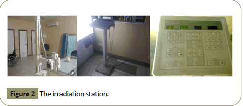
The radiographs were obtained using the same experimental procedures. All white lights in the darkroom were switched off and only the safelight was on during processing. An unexposed film in total darkness was placed in a light-tight cassette containing intensifying screens. In order to obtain the radiograph, the phantom was mounted in the direction of the X-ray beam with the film placed behind the object. The films were marked using markers. To obtain a good quality film when processing the intensity of the safe light is being checked (Figure 2).
The decision as to the length of exposure, kV and mAs was made by the Radiographer on the basis of previous experience of similar examinations.
Basically, the exposure selection in this experiment was based on the aim of the work. After proper collimation and preselection process at the control panel, exposure was made on the control knobs and the incident divergent beam traverses the compression plate, through the specimen and film, conveying part of their energy to the screen phosphor which produces the light that exposes the film leading to the formation of the latent image. Exposed films were passed through the hatch into the darkroom where the cassettes were open in the darkness. The films were then processed under fixed processing factors. While for the absorbed dose, the dose meter was placed in front of the constructed skull at the center in line with the sensor at the center of the calumniated beam in order to measure the input direct radiation and also for the output the same procedure but with the dose meter placed behind the constructed skull to measure the output dose.
Determination of suitable exposure factors
A suitable exposure factors range for the skull X-ray was determined using the Perspex phantom. This was done by X-raying it at a fixed Focal Film Distance (FFD) of 120 cm under varying exposure factors so as to get the least- exposure factors that can produce a quality image.
Image quality assessment and determination
An evaluative panel of three experienced staff consisting of one Radiologist, one Imaging Scientist, and one Physicist was used to assess the images of the Perspex phantom and determine the clear and quality ones. All of these individuals underwent appropriate training prior to any evaluation of the images.
Effects of exposure factors on skull radiographs
The determination of the effects of exposure factors was done by X-raying the phantom at varying factors as follows:
• Varying Kilovolts: This was done at a fixed FFD of 120 cm and constant mAs of 50 mAs.
• Varying milli Ampere seconds: This was done at a fixed FFD of 120 cm and constant kilo Voltage of 81 kV.
Calculation of skull radiation dose absorbed
This was calculated using the formula derived from the definition of absorbed dose which is given mathematically as:
Absorbed dose=Input dose - Output dose
(I.e. the different between the input and output dose)
Ad = Id −Od
Where
Ad= Absorbed dose
Id=Input dose
Od=Output dose
(The unit is mGy).
Results and Discussion
Assessment on the quality of the skull radiographs
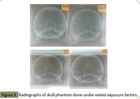
The image qualities of the radiographs are assessed according to the radiological and anatomical classification, and the following were discovered (Figure 3).
From Table 1 and Table 2 below, the results show that films with 81 kV and 32 mAs are the best image quality images of the examination. This is because the sharpness, contrast and clarity of the films made it possible to see clearly the detailed anatomical features of the skull. And also show us that as kV increases, the radiograph quality loses out. Also as mAs increases, the radiograph quality loses out [13,14].
| Kv | Image quality grade |
|---|---|
| 81 | 1 |
| 85 | 2 |
| 90 | 3 |
| 96 | 4 |
| 102 | 5 |
Table 1: Radiographic quality under varying kV.
| mAs | Image quality grade |
|---|---|
| 32 | 1 |
| 40 | 2 |
| 50 | 3 |
| 63 | 4 |
| 80 | 5 |
Table 2: Radiographic quality under varying mAs.
Suitable exposure factors determination
From Tables 1 and 2, the suitable exposure factors that can be used to obtain a quality skull X-ray radiograph is as follows:
1. 81-85 potential difference in kilovolts (kV) and
2. 32-40 tube load (mAs)
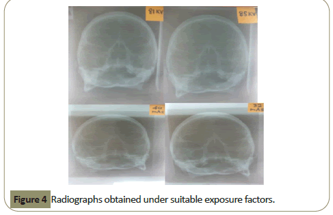
Figure 4 shows a radiograph obtained from the suitable exposure factors, which is in line with the International Commission on Radiation Units and measurement (ICRU) standard.
Effects of exposure factors on the quality of skull x-ray radiography
To observe the effects of the exposure factors on skull X-ray the constituents of the exposure factors (kilo Voltage "kV",milli Ampere seconds “mAs”) were varied one after the other as the skull phantom was exposed. The fixed factors used are the optimum factors used for skull X-rays exanimation for the research.
Effects of Kilo Voltage (kV)
In this experiment, the mAs and FFD were kept constant while the kV was varied as shown below (Table 3):
| Film No. | FFD (cm) | kV | mAs | Input Dose (mGy) | Output dose (mGy) | Absorbed Dose (mGy) |
|---|---|---|---|---|---|---|
| F1 | 120 | 81 | 50.0 | 2.64 | 0.285 | 2.355 |
| F2 | 120 | 85 | 50.0 | 2.97 | 0.367 | 2.603 |
| F3 | 120 | 90 | 50.0 | 3.29 | 0.455 | 2.835 |
| F4 | 120 | 96 | 50.0 | 3.68 | 0.551 | 3.129 |
| F5 | 120 | 102 | 50.0 | 4.08 | 0.623 | 3.457 |
Table 3: Results obtained under varying potential difference in kilovolts (kV).
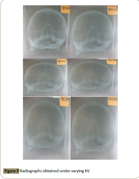
Figure 5 Shows that the contrast of the film was affected by varying the kV. This is because the kV affects the energy of the X-ray beam. As the voltage increases, it tends to increase the acceleration of the electrons travelling to the target, which also increases the energy of the X-rays produced. By so doing, harder X-rays will be produced which increases the penetration power and reduces the attenuation level of the tissues of the skull [14].
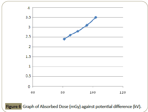
Figure 6 shows as the kV increases, the absorbed dose increases but not directly proportional.
Effect of milli-Ampere seconds (mAs)
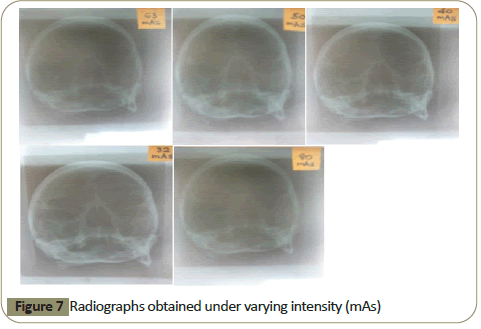
Here the exposure FFD (cm) and the kilo Voltage (kV) were kept constant and the milli-Ampere seconds (mAs) was varied from 32-80 mAs as shown in below (Table 4) (Figure 7).
| Film No. | FFD (cm) | kV | mAs | Input Dose (mGy) | Output dose (mGy) | Absorbed Dose (mGy) |
|---|---|---|---|---|---|---|
| F1 | 120 | 81 | 32 | 1.75 | 0.299 | 1.451 |
| F2 | 120 | 81 | 40 | 2.23 | 0.419 | 1.811 |
| F3 | 120 | 81 | 50 | 2.82 | 0.481 | 2.339 |
| F4 | 120 | 81 | 63 | 3.50 | 0.700 | 2.800 |
| F5 | 120 | 81 | 80 | 4.44 | 0.937 | 3.503 |
Table 4: Results obtained under varying intensity or tube load (mAs).
Figure 4 show that as the mAs increases the darkening of the film also increases. This means that the mAs determine how much current is allowed to flow through the filament which is in the cathode side of the tube. The effect of mAs is not linear in as much as increasing the number of X-rays produced is done by increasing the mAs and vice-versa which has affected the darkening of the radiographs [15].
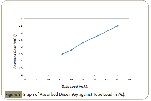
Figure 8 shows as the mAs increases, the absorbed dose increases but not directly proportional.
Calculated absorbed dose obtained
The result of the calculated absorbed dose using equation 14 shows that:
This was calculated using the formula derived from the definition of absorbed dose which is given mathematically as:
Absorbed dose=Input dose - Output dose
(I.e. the different between the input and output dose)
Ad = Id −Od
Where
Ad=Absorbed dose
Id=Input dose
Od=Output dose
(The unit is mGy).
In Table 3 where the kV was varied, we observed that the absorbed dose increased as we increase kV. This is because the energy of the X-rays increased with increase in kV, thereby causing more hard X-rays to penetrate through the phantom which lead to the increase in absorbed dose [16].
In Table 4 where the mAs were varied, we observed that the absorbed dose increased as we increase mAs. This is because the intensity (number of electrons) of the X-rays increased with increase in mAs thereby causing more (electrons) X-rays to be administered to the phantom which lead to the increase in absorbed dose [17].
In general, an increase in energy and intensity increases the dose absorbed by the patient.
The study also shows that suitable exposure factors can substantially reduce patients’ doses while maintaining acceptable image quality with no additional cost implication. From the results presented we conclude that the suitable exposure factors for skull X-rays are within the range: 81 – 85 kV, 32- 40 mAs.
Conclusion
To A steady increase in professional and public concern regarding the biological effects of ionizing radiation on human tissues motivated us to undertake this project. There is a need for radiography professionals to improve imaging techniques with minimal radiation exposure to patients at all times. The data (values) obtained in this study (skull X-ray) shows that exposure factors affect the total image quality and the absorbed dose received by the patient. Therefore, the Radiographer has to come to a compromise between the image quality and the radiation dose to the patient according to ALARA (As Low as Reasonably Achievable) principle. The findings of this work can be used as reference values for skull X-ray procedures in radiological establishments.
Recommendations
• This work should be put in practice to reduce doses to the patients’ general public and the staff of the department since it is easy to apply and of no cost implication.
• Research should be carried out to find the suitable exposure factors of X-rays in other parts of the body such as pelvis, shoulder, abdomen, foot and so on.
• Further research should be carried out separately for both sexes and for different ages to see if the exposure factors differ.
References
- Geijer H (2001) Radiation dose and image quality in diagnostic radiology: optimization of the dose-image quality relationship with clinical experience from scoliosis radiography, coronary intervention and a flat-panel digital detector. 1-76.
- Brenner D, Huda W (2008) Effective dose: A useful concept in diagnostic radiology? Radiat Prot Dosimetry. 128(4): 503-508.
- Podgorsak EB (2005) Radiation oncology physics. 92(6): 158-176.
- Armstrong P, Wastle (1987) Diagnostic Imaging (2nd edn) 198-204.
- Mengistu TM, Sprawls (1993) Physics Principles of Medical Imaging (2nd edn) 165-216.
- Beir (2007) Health Risks from exposure to low levels of ionizing Radiation, Board on radiation effects Research VII phase. 267-312.
- Hay A , Hughes (1986) D First Year Physics for Radiographers (3rd edn)139-247
- Chesney DN, Chesney MO (1984) Radiographic Imaging (4th edn) 291-329.
- Dingson (2006) Effect of some exposure factors on the film density of X-ray radiographic films unpublished project. 31-33.
- Fulea D, Cosma C, Pop IG (2009) Monte Carlo method for radiological X-ray examinations. Romanian Journal in Physics. 54(7-8):629-639.
- Meredith, WJ, Massey (1986) Fundamental Physics of Radiology (3rd Edn) 57-73.
- Armstrong (1990) National Radiological Protection Board (NRPB) Royal College of Radiologist. 120-125.
- Olusola OI, Bassey CE, Vincent UE (2005) Dose determination in radiodiagnosis using exposure factors. Nigerian Journal of Physics. 17(2): 245-250.
- Palmer PE (1995) Manual of diagnostic ultrasound. World Health Organization. 9: 2-10
- Sutton (1995) Radiology and Imaging for Medical Students (6th Edn) 217-226.
- Nwankwo (2004) Determination of Exposure factors on the quality of skull X-ray radiographs. A Thesis of Physics Department University of Jos. 30-35
- Tsapaki V, Tsalafoutas IA, Chinofoti I, Karageorgi A, Carinou E, et al. (2007) Radiation doses to patients undergoing standard radiographic examinations: a comparison between two methods. The British Journal of Radiology.80(950): 107-12.
Open Access Journals
- Aquaculture & Veterinary Science
- Chemistry & Chemical Sciences
- Clinical Sciences
- Engineering
- General Science
- Genetics & Molecular Biology
- Health Care & Nursing
- Immunology & Microbiology
- Materials Science
- Mathematics & Physics
- Medical Sciences
- Neurology & Psychiatry
- Oncology & Cancer Science
- Pharmaceutical Sciences

