Estimation of Radiation Dose in the Neonatal Intensive Care Unit (NICU)
Emadeldin B, Abukonna A
1King Saud University, (KSA), Riadh
2Sudan University of Science and Technology, College of Medical Radiologic Science, Khartoum, Sudan
- *Corresponding Author:
- Abukonna A
Sudan University of Science and Technology
College of Medical Radiologic Science
Khartoum, Sudan
Tel: 0024983771818
E-mail: konaa17@hotmail.com
Received date: April 27, 2016; Accepted date: May 16, 2016; Published date: May 22, 2016
Citation: Emadeldin B, Abukonna A. Estimation of Radiation Dose in the Neonatal Intensive Care Unit (NICU). Insights Med Phys. 2016, 1:2
Abstract
An experimental study was carried out to determine the radiation doses received by the brain, thyroid glands and the genital organs (i.e. ovaries testicles) of the babies in the Neonatal Intensive Care Unit (NICU) during chest radiography. A Perspex block phantom of similar size to the neonate was exposed to radiation using the actual average exposure parameters that applied normally in the NICU. The doses were measured using thermo-luminance lithium fluoride dosimeter chips (TLD). The entrance and exit doses were then measured for the brain, thyroid and gonads organs. The measurement was obtained with and without shielding. The results of our study showed that infants did not receive what might be considered excessive radiation from diagnostic modalities. Entrance Skin Dose (ESD) was found to be below the European Committee (EC) reference dose of 80 mGy for mobile chest radiographs. Applying the radiation protection shield such as 0.5 mm lead rubber sheet is of great value in reducing the radiation doses to the brain cells and the gonadal organs.
Keywords
ESD, Absorbed dose, Neonatal Intensive Care Unit (NICU)
Introduction
Radiography is one of the important new mean of investigation in modern medicine. Diagnostic radiology is increasingly used in the assessment and treatment of neonates requiring intensive care [1]. Therefore, in a department such as the Neonatal Intensive care Unit (NICU), it becomes of a great importance as it gives the physician an easy and accurate way of examining neonate. The commonest clinical indications of chest radiography on NICU include; respiratory distress syndrome (RDS), acquired pneumonia pre/ post-delivery and pneumothorax which is a complication of RDS or aspiration ventilation in the newborn. Consequently there are a lot of risks in using x-rays on the human body cells. These risks can be divided into somatic and genetic effects. The somatic effects include infertility, cancer and cataracts. The genetic effects show abnormal features in the infants after the excessive exposure of the pregnant mother to radiations [2]. However in an NICU, where the incubator plays the role of the mother womb, it becomes impossible to avoid radiation that is because the neonate is got to have an x-ray taken at least to justify his /her stay in the unit, where the premature newly born sick children infants are kept to be looked after [3].
In the NICU, where physicians weigh the risks and benefits of using x-rays as investigation tool. They tend to go for it as they are in a condition of life saving. Nevertheless, thinking of those risks associated with using of x-rays motivated researches to find a way to keep them to the lowest level that is possible.
Radiographic examinations of neonates are particularly critical because of delayed radiogenic cancers as a consequence of a relative longer life expectancy [1]. The small size of neonates, especially of premature infants, places all organs within the useful beam, resulting in a higher effective dose per radiograph than may be the case with older children and adults. Therefore, radiation doses for neonatal X-ray examination should be kept to a minimum. It is also important to ensure that radiation doses from repeated radiographic examinations carried out frequently in the same room of the NICU should be kept at a minimum. The aim of this work was to determine the radiation doses received by the infants from radiographic exposure in order to find the dose received by brain, thyroids and gonadal organs (i.e. ovaries and testicles) during an antero-posterior chest x-ray with and without shielding.
Method
Experimental design
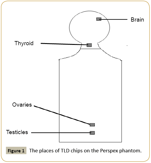
A Perspex block phantom (35 cm trunk length, 10cm trunk width and 10 cm thickness) was used to simulate the size of the neonate and was exposed to the actual average exposure parameters that usually used in the NICU. Thermo-luminance lithium fluoride chips were used to determine the radiation dose readings. To ensure high specificity and sensitivity of the study dose calculations, the room pressure and temperature were measured. The pressure was equal to 944.6mbar and the temperature was equal to 20.7°C. The TLDs supposed to be used in experiment were calibrated to read to a level close to that assumed to be gotten from the experiment as follows: Three different exposures were made at exactly, same distance between the x-ray tube and a 37D Pitman dosimeter. The three points of readings were used to plot a graph later on for more sensitivity and reliability of dose measurement calculations done by the medical physicist.
The exposure factors that normally used for chest in the RK Hospital were checked and the mean values were taken (66 kvp, 0.5 mAs and 110 cm as Focal to Image receptor Distance).
The TLDs were then used on three different regions (Figure 1). The experiment was done twice; one with lead shielding (0.5 mm lead rubber sheets on the edge of the phantom) and the other without shielding, thirty exposures were made and the dose per exam was calculated.
Results and Discussion
Chest x- ray detect a very frequency of pulmonary abnormalities, significant interval changes, and tube and catheter malposition [4]. Radiograph of neonate has a significant impact on decision concerning management of patient despite numerous radiographs, i.e. x-rays, produced for each neonate. The entrance skin dose, the exit skin dose and the absorbed dose for the brain, thyroid and the gonadal organs (ovaries and testicles) were calculated (Tables 1 and 2).
| Body Organ | Entrance Skin Dose "mGy" ESD |
Exit Dose "mGy" |
AbsorbedDose "mGy" |
|---|---|---|---|
| Brain | 4.35 | 2.9 | 1.45 |
| Thyroid | 17.69 | 5.51 | 12.18 |
| Ovaries | 11.31 | 6.67 | 4.64 |
| Testicles | 8.41 | 1.16 | 7.25 |
Table 1: The dose readings when no lead shielding is applied (Dose/ Exam).
| Body Organ | Entrance Skin Dose "mGy" ESD |
Exit Dose "mGy" |
AbsorbedDose "mGy" |
|---|---|---|---|
| Brain | 2.32 | 1.74 | 0.58 |
| Thyroid | 15.08 | 4.64 | 10.44 |
| Ovaries | 4.35 | 3.77 | 0.58 |
| Testicles | 2.9 | 0.58 | 2.32 |
Table 2: The dose readings when lead shielding is applied.
The results of our study show that infants did not receive what might be considered ‘‘excessive’’ radiation from diagnostic modalities. ESDs were found to be below the EC reference dose of 80 mGy for mobile chest radiographs and below the National Radiological Protection Board (NRPB) reference dose of 50 mGy for chest examination [5,6]. These doses are for a single examination only and therefore they could be higher if more chest x-rays were made as is the case in the NICU where neonate might need to stay for a long period of time. This because the radiation dose effects are cumulative. i.e., radiation dose never get to zero or disappear.
Comparing the readings of both tables, there was a significant difference between doses when applying the lead shielding; Because of their sizes, a relatively large area of the infants was irradiated and it was more difficult to shield radiosensitive organs. In addition, measurement of infant dose must be performed in sterile conditions as soon as possible because of the high sensitivity of the infants to infection. Applying the radiation protection measures such as 0.5 mm lead rubber sheets are of great value in reducing the radiation doses to the brain cells and the gonadal organs. However, the thyroids are still getting very high dose even when applying the shielding. The only way to try to reduce the radiation dose is by applying a very well collimated x-ray beam.
The results show the risk from neonatal radiation to be fairly low, and it is considered to be substantially outweighed by the clinical benefit of the radiograph in assessing the progress of a sick baby. However, the risk vs. benefit of each radiograph is important and must be considered carefully, especially as radiation effects are cumulative.
References
- Olgar T, Onal E, Bor D, Okumus N, Atalay Y, et al. (2008) Radiation exposure to premature infants in a neonatal intensive care unit in Turkey. Korean J Radiol 9: 416-419.
- Streffer C , Shore R, Konermann G, Meadows A, Uma Devi P, et al. (2003) Biological effects after prenatal irradiation (embryo and fetus). A report of the International Commission on Radiological Protection. Ann ICRP 33: 5-206.
- Bahreyni Toossi MT, Malekzadeh M (2012) Radiation dose to newborns in neonatal intensive care units. Iran J Radiol 9: 145-149.
- Sivit CJ, Taylor GA, Hauser GJ, Pollack MM, Bulas DI, et al. (1989) Efficacy of chest radiography in pediatric intensive care. AJR Am J Roentgenol152: 575-577.
- Ropolo R, Isoardi P, Catuzzo P, Bianchi SD, Bradac GB, et al.(2001) Check of compliance with quality criteria for diagnostic radiographic images in quality control. Radiol Med101: 75-81.
- Armpilia CI, Fife IA, Croasdale PL (2002) Radiation dose quantities and risk in neonates in a special care baby unit. Br J Radiol 75: 590-595.
Open Access Journals
- Aquaculture & Veterinary Science
- Chemistry & Chemical Sciences
- Clinical Sciences
- Engineering
- General Science
- Genetics & Molecular Biology
- Health Care & Nursing
- Immunology & Microbiology
- Materials Science
- Mathematics & Physics
- Medical Sciences
- Neurology & Psychiatry
- Oncology & Cancer Science
- Pharmaceutical Sciences

